A ERA synopsis for nephrology practice of the 2023 ESH Guidelines for the Management of Arterial Hypertension
NDT Kidney Art Gallery
Welcome to NDT's Kidney Art Gallery
Welcome to NDT’s Kidney Art Gallery, a captivating visual journey into the intricate world of the kidneys!
This collection of stunning images submitted by NDT’s most creative readers and artists offers a unique glimpse into the complex structures and functions of the kidneys, showing the remarkable interplay of form and function in these vital organs. We invite you to explore the fascinating realm of the kidneys and see kidney health and disease from a new perspective.
Selected images will appear on the covers of NDT:
12/76 elements

Author: Anil Kumar Saxena, Dubai
Digital art
Info:
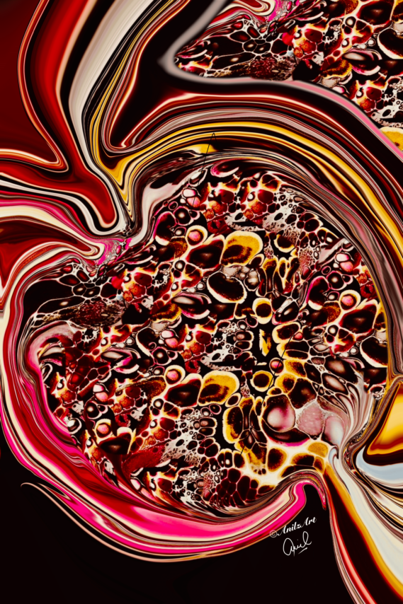
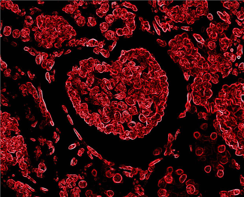
This artwork presents a stylized representation of glomeruli in an abstract, minimalistic form. Bright red strokes evoke the intricate vascular structures, swirling dynamically against a dark background.
The fluidity of the curves, contrasted with sharp lines, symbolizes the delicate balance of function and form in the microscopic kidney filters.
From:
Anil Kumar Saxena, Dubai

Author: Anil Kumar Saxena, Dubai
Digital art
Info:
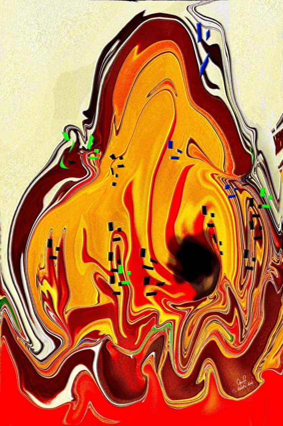
The artwork represents an abstract depiction of an inflamed podocyte, which is a type of cell in the kidney involved in the filtration process. The vibrant red, yellow, and orange tones symbolizes inflammation.
From:
Anil Kumar Saxena, Dubai

Author: Anil Kumar Saxena, Dubai
Digital art
Info:
The vibrant chaos of life splinters in a flash—dreams splattered across a jagged landscape of shock and despair. The news of the end stage kidney disease and dialysis dependence pierces the soul like molten streaks, crimson and gold, swirling with the black void of uncertainty. The delicate balance shattered, leaving veins of hope entangled in dark realization.
Yet within the turbulence, the fight begins; the will to endure finds form among the chaos, weaving fragile, glistening strands of resilience.
From:
Anil Kumar Saxena, Dubai

Author: Anil Kumar Saxena, Dubai
Digital art
Info:
This vivid artwork symbolizes the relentless work of the glomeruli, filtering blood day and night. The swirling blues, pinks, and reds mirror the constant flow of life, with each curve and wave representing filtration, absorption, and secretion. The abstract forms convey the ceaseless activity within the kidneys, their colors reflecting oxygenated and deoxygenated blood, symbolizing balance and rhythm.
It is a tribute to the glomeruli’s tireless duty in sustaining life’s inner harmony.
From:
Anil Kumar Saxena, Dubai

Author: Anil Kumar Saxena, Dubai
Digital art
Info:
The fetus nestled within the Renal outline radiates an ethereal glow, symbolizing hope born from adversity. It is a testament to the miracle of creation, where, even in the shadow of illness, the promise of renewal prevails. The swirling hues reflect the turbulence and warmth of this journey—a vivid gesture of survival, defying limits with grace. “Germination” embodies the profound triumph of life against the odds, where resilience blossoms within the most fragile vessel. (Inspired by the real-life experience when a young lady with ESRD due to lupus nephritis on dialysis delivered a healthy baby through C-section)
From:
Anil Kumar Saxena, Dubai

Author: Lars Koch & Gabriela Jiang, Germany
Digital art
Info:
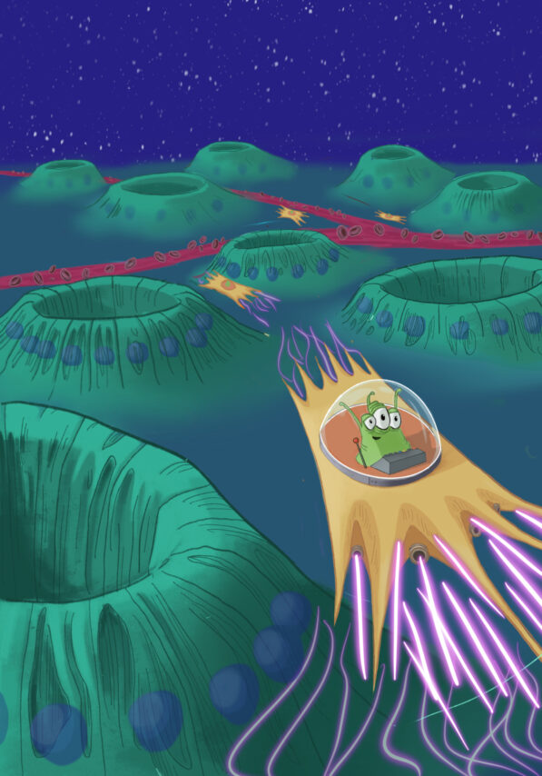
The proposed cover art shows an abandoned moon landscrape with several craters representing kidney tubuli. Aliens are moving in their spaceships, representing myofibroblasts, in between the craters (in the interstitium) and fire around extracellular matrix. The cover art aims to explain basic principles of kidney fibrosis in an engaging way.
From:
Lars Koch & Gabriela Jiang, Germany

Author: Nuno Moreira Fonseca & David Navarro, Portugal
Urine & urine microscopy
Info:
Struvite crystals, bacteria and leucocytes (phase contrast, 400x)
From:
Nuno Moreira Fonseca & David Navarro, Portugal

Author: Nuno Moreira Fonseca & David Navarro, Portugal
Urine & urine microscopy
Info:
Monohydrate calcium oxalate crystals (phase contrast, 400x)
From:
Nuno Moreira Fonseca & David Navarro, Portugal

Author: Nuno Moreira Fonseca & David Navarro, Portugal
Urine & urine microscopy
Info:
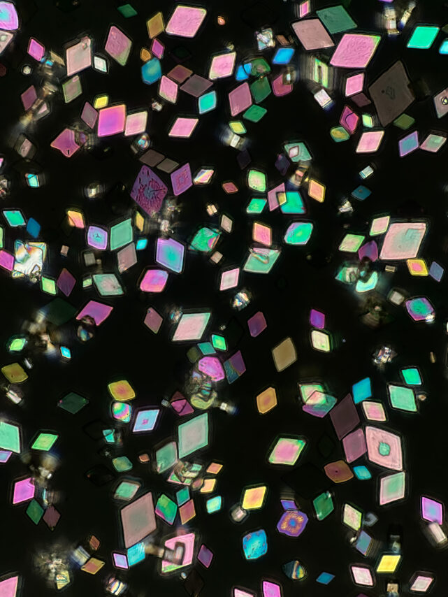
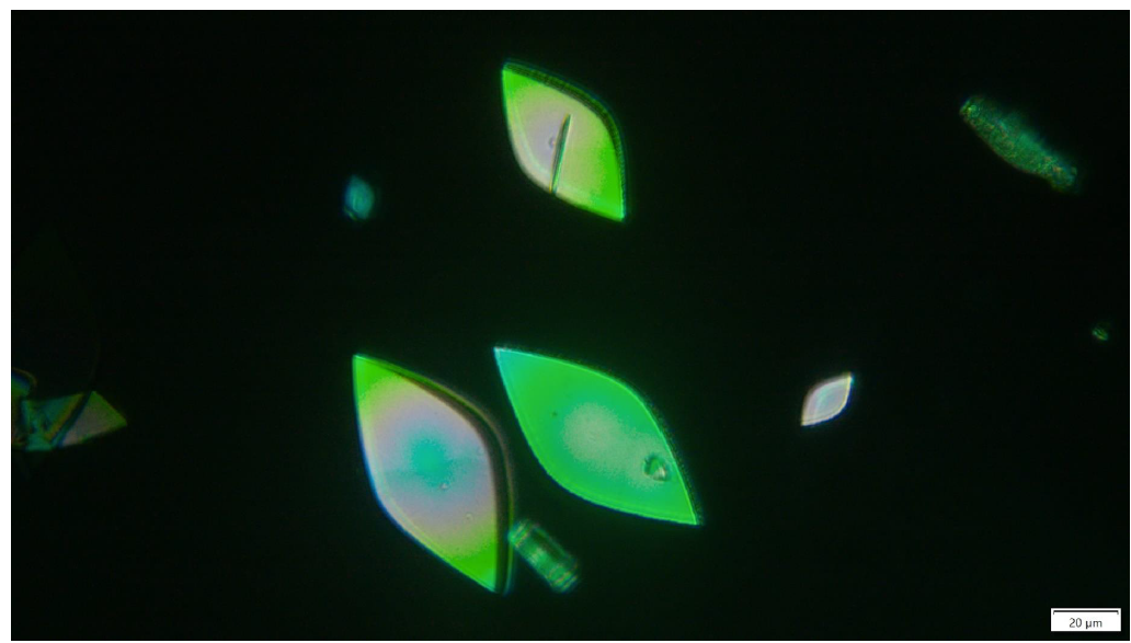
Uric Acid Crystals (polarized light, 160x)
From:
Nuno Moreira Fonseca & David Navarro, Portugal
Author: Nuno Moreira Fonseca & David Navarro, Portugal
Urine & urine microscopy
Info:
Uric Acid Crystals (polarized light, 400x)
From:
Nuno Moreira Fonseca & David Navarro, Portugal

Author: Taner Çamsarı, Türkiye
Info:
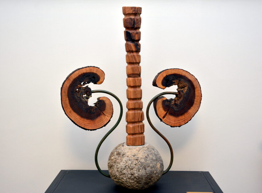
Kidney sculpture made of granite, copper, olive wood and Judas tree wood
From:
Taner Çamsarı, Türkiye

Author: Mayleen Laico, Philippines
Digital art
Info:
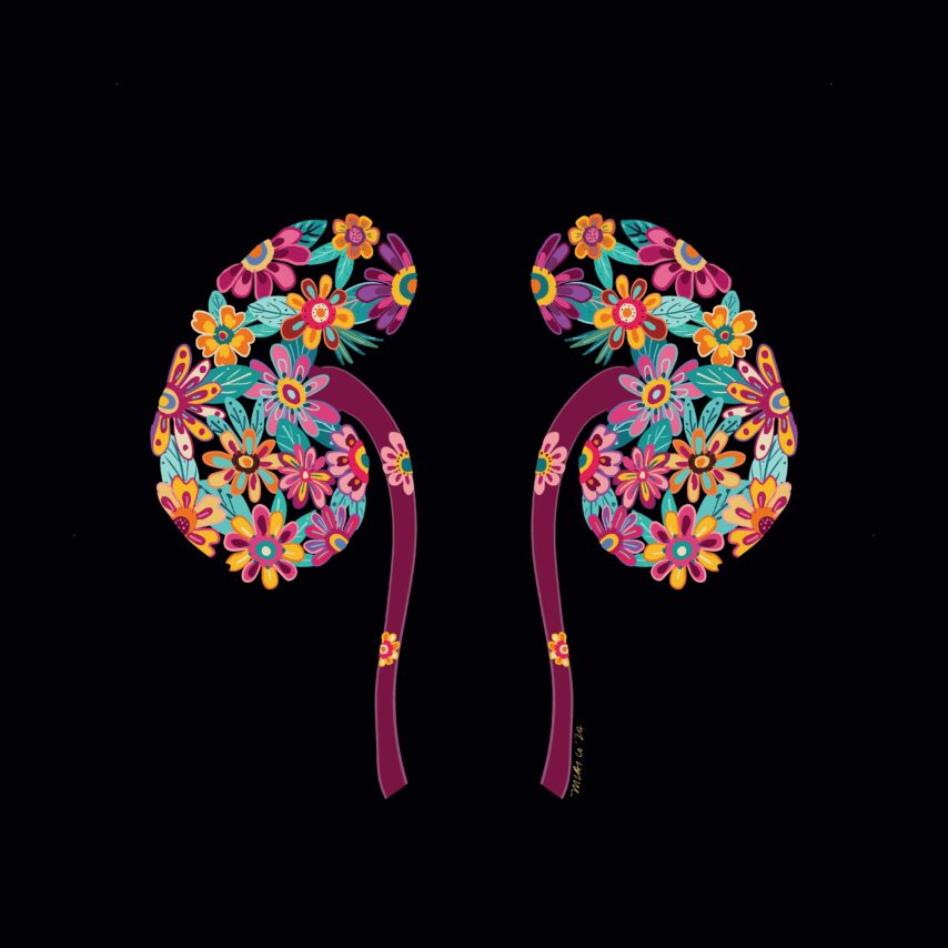
The title is “Celebration”. Celebrating small victories in kidney care.
From:
Mayleen Laico, Philippines
Author: Swetha Sriramoju, USA
Drawing
Info:
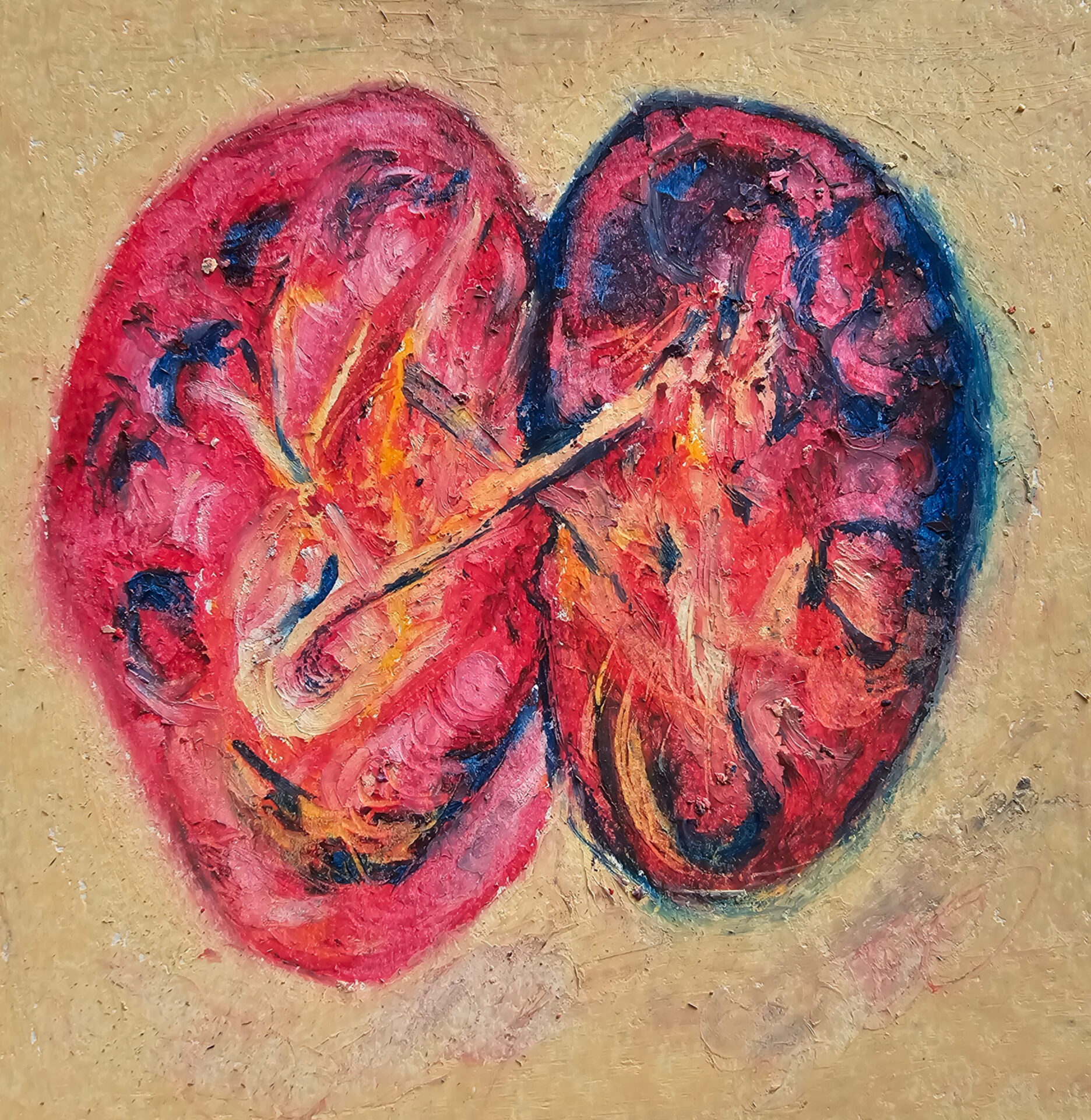
Artwork entitled, Gross Anatomy of Kidneys, was drawn in oil pastel to illustrate studying the anatomy of the kidneys.
From:
Swetha Sriramoju, USA

Author: Oliver Kretz, Germany
Digital art
NDT Cover October 2024
Info:
Nephros – panta rhei
From:
Oliver Kretz, Hamburg, Germany
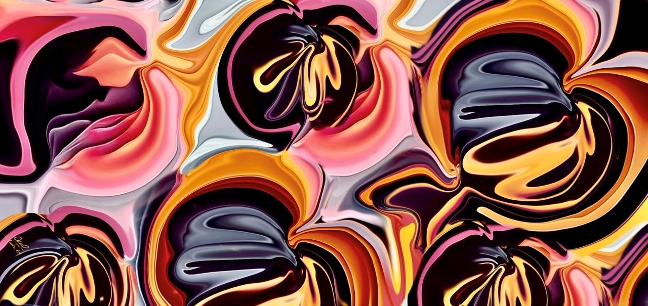
Author: Anil Kumar Saxena, Dubai
Digital art
Info:
A Beautiful Family of Glomeruli
From:
Anil Kumar Saxena, Dubai

Author: Nisrine Bennani Guebessi, Morocco
Histology
Info:
The image displays glomerular capillary lumens filled with large lymphomatous cells, indicative of intravascular large B-cell lymphoma. Masson’s trichrome stain at 200x magnification.
From:
Nisrine Bennani Guebessi, Morocco
Pathologist and Professor of pathology, department of pathology CHU IBN ROCHD, Casablanca
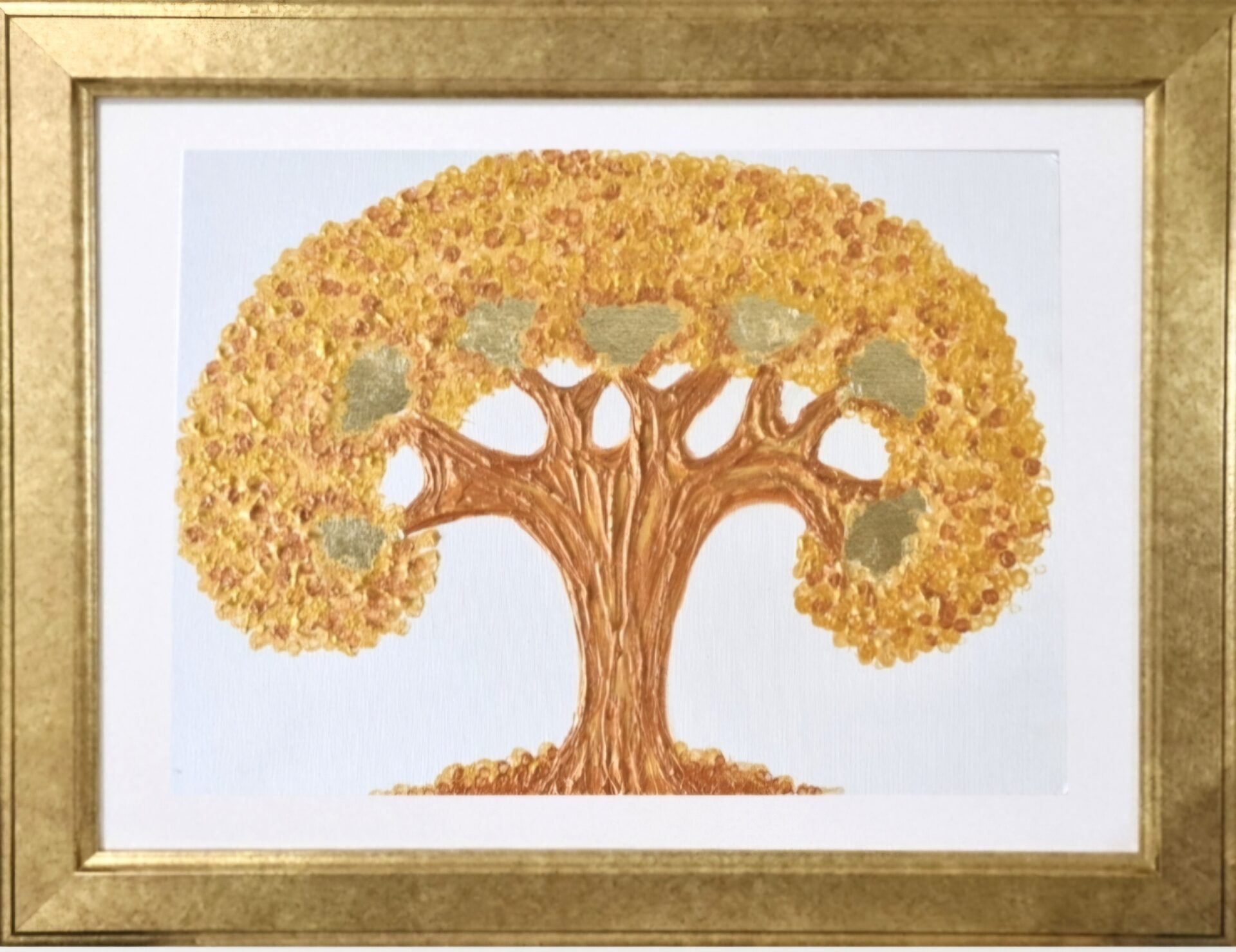
Author: Ailene Buelva-Martin, Philippines
Drawing
Info:
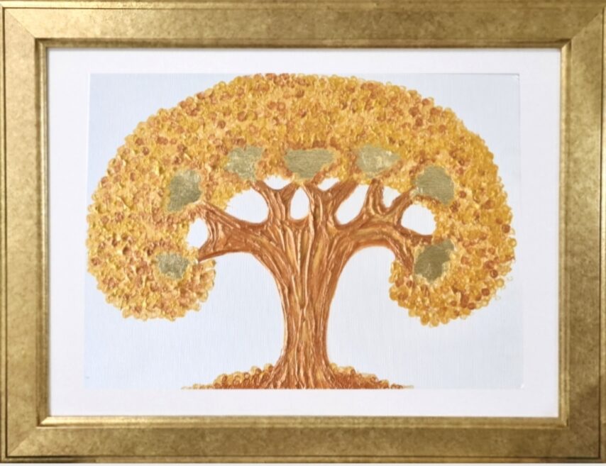
The Golden Kidney Tree. Acrylic on canvas with gold foil.
From:
Ailene Buelva-Martin, Philippines

Author: Mayleen Laico, Philippines
Drawing
Info:
The title of the painting is “Stained”. It is inspired by the immunoflourescent staining of glomeruli done in the histopathologic evaluation of glomerular disease.
From:
Mayleen Laico, Philippines

Author: Nicole Endlich, Germany
Advanced microscopy
Info:
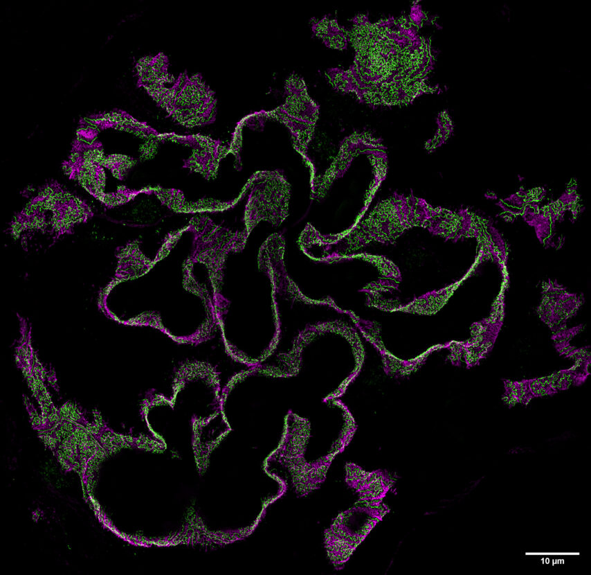
Human glomerulus (podocin in green, synaptopodin in magenta)
From:
Nicole Endlich, Germany

Author: Maximilian Schindler, Germany
Advanced microscopy
NDT Cover August 2024
Info:
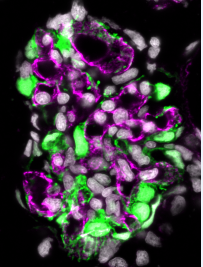
This confocal image shows a cryosection of a larval zebrafish glomerulus at 5 days post fertilization. The endogenously expressed eGFP under control of the nphs2 promotor labels podocytes in green, including the major processes. Magenta depicts the glomerular endothelial cells with an antibody against Ehd3.
From:
Maximilian Schindler, Germany
Department of Anatomy and Cell Biology, University Medicine Greifswald, Nicole Endlich’s Lab
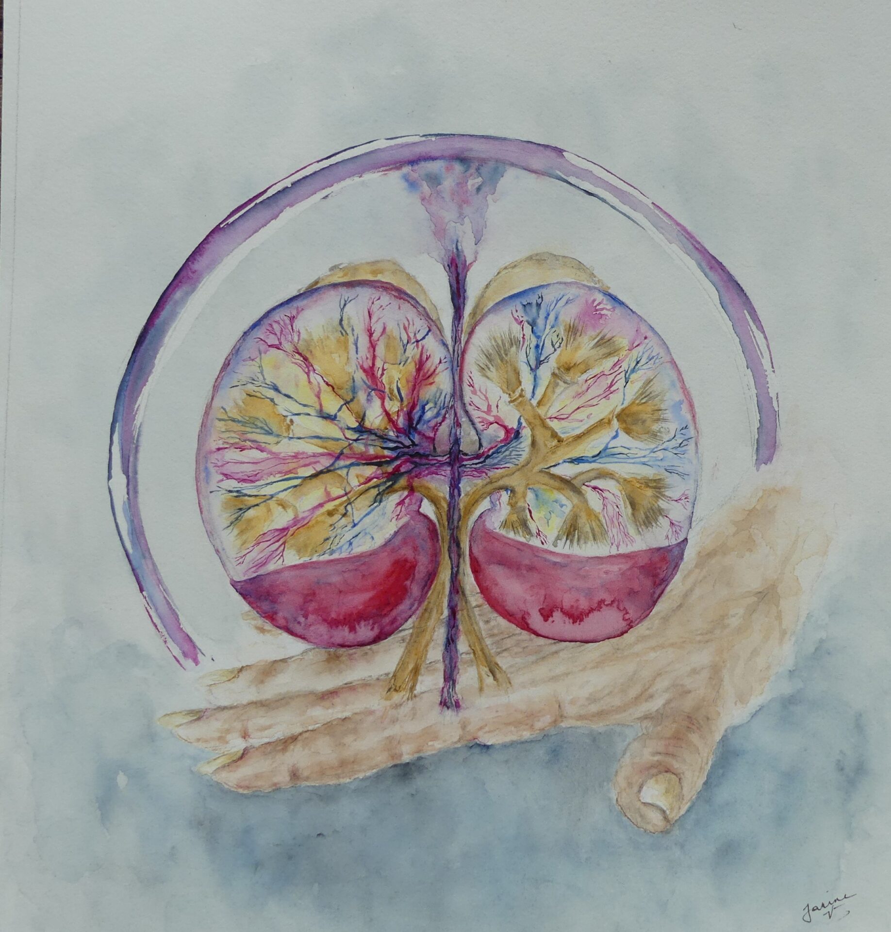
Author: Jeanine Verbeke, Belgium
Drawing
Info:
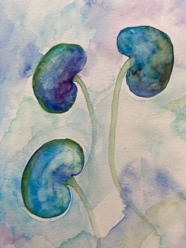
Aquarelle painting called ‘Care for your Kidneys’.
From:
Jeanine Verbeke, Belgium

Author: Marieta Theodorakopoulou, Greece
Imaging
NDT Cover June 2024
Info:
Combination of (i) immunofluorescence images stained for IgA and (b) renal magnetic resonance angiography images, in a patient with IgA and atherosclerotic renovascular disease (ARVD)
From:
Marieta Theodorakopoulou, Greece
1st Department of Nephrology, Hippokration Hospital, Aristotle University of Thessaloniki, Thessaloniki

Author: Margaux Van Wynsberghe, France
Advanced microscopy
Info:
Glomerulus lover – Representative image showing immunofluorescent staining for nephrin (green), FAK (red) and DAPI (blue) on a piece of human nephrectomy.
From:
Margaux Van Wynsberghe, France
IMRB Inserm U955, Hôpital Henri Mondor
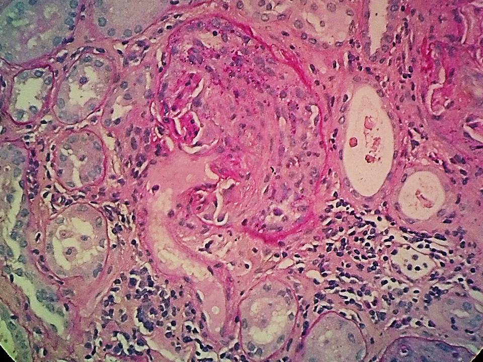
Author: Panagiotis Pateinakis, Greece
Histology
Info:
“Inception”- like, kidney-shaped nephron with a cellular crescent, from a patient with pauci-immune ANCA-associated renal vasculitis
From:
Panagiotis Pateinakis, Greece
Nephrology Department, “Papagergiou” General Hospital
Author: Maria Lambropoulou, Greece
Histology
Info:
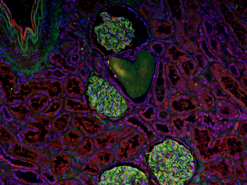
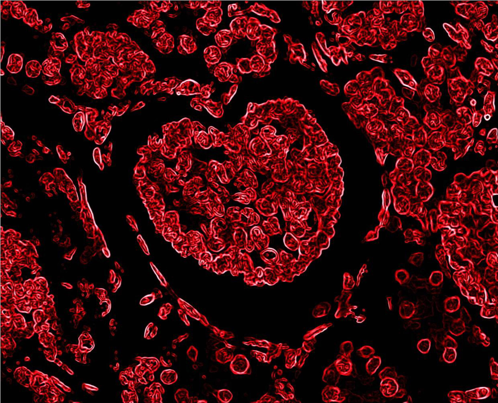
Kidney histological section in which a glomerulus forming the most iconic symbol of love; a big heart has been discovered in a hematoxylin-eosin slide (negative colors) after a hard day working on microscope
From:
Maria Lambropoulou, Greece
Pathologist & Professor of Histology-Embryology
Medical Department, Democritus University of Thrace, Alexandroupolis

Author: Maria Lambropoulou, Greece
Histology
NDT Cover May 2024
Info:
Kidney histological section in which the “heart” represents collecting tubule (hematoxylin-eosin staining, 200×)
From:
Maria Lambropoulou, Greece
Pathologist & Professor of Histology-Embryology
Medical Department, Democritus University of Thrace, Alexandroupolis

Author: Mayleen Laico, Philippines
Digital art
NDT Cover April 2024
Info:
The Gift of Organ Donation
From:
Mayleen Laico, Philippines
Author: Oliver Kretz, Germany
Electron microscopy
Info:
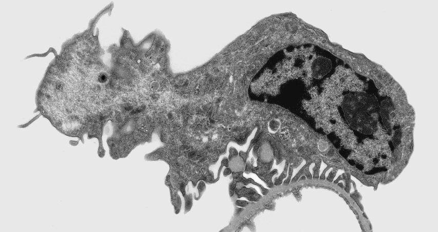
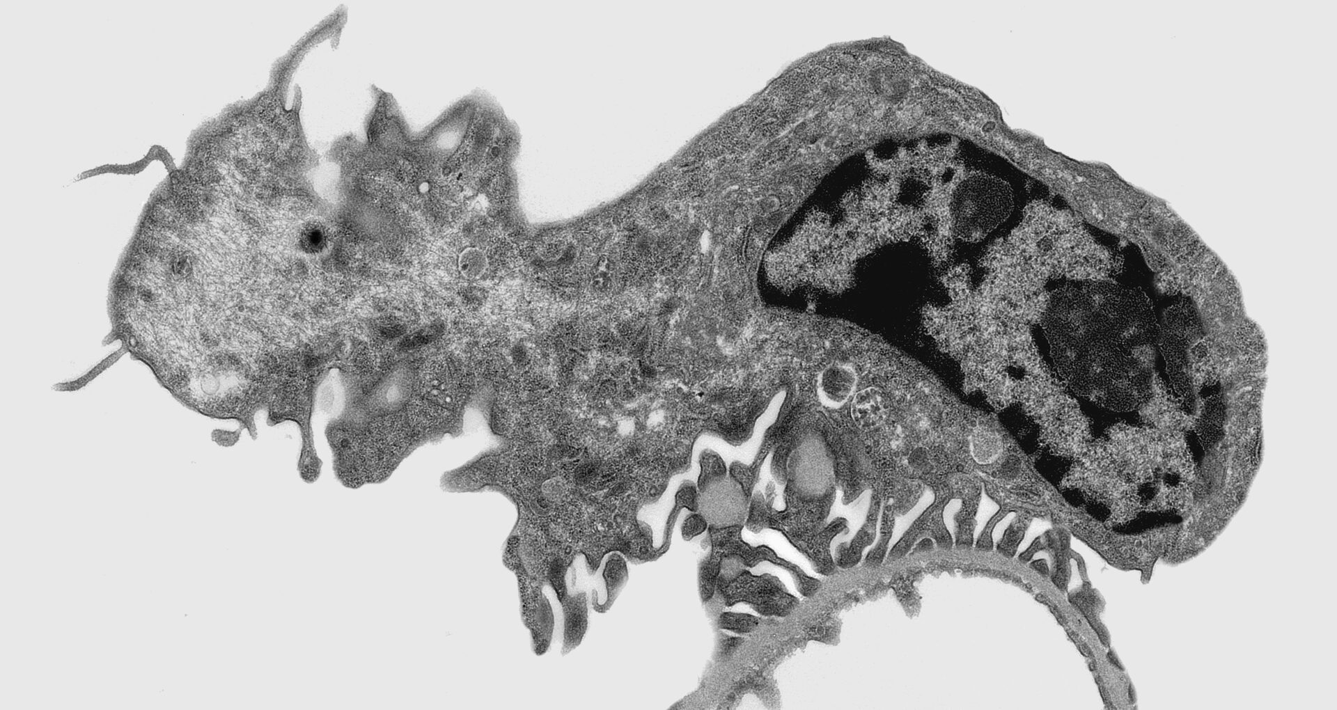
Transmission electron microscopy image of the murine renal filtration barrier. Random sections of the podocytes are sometimes reminiscent of prehistoric creatures.
From:
Oliver Kretz, Germany
Author: Yaoxian Xu | Rafael Kramann, Germany
Cells
Info:
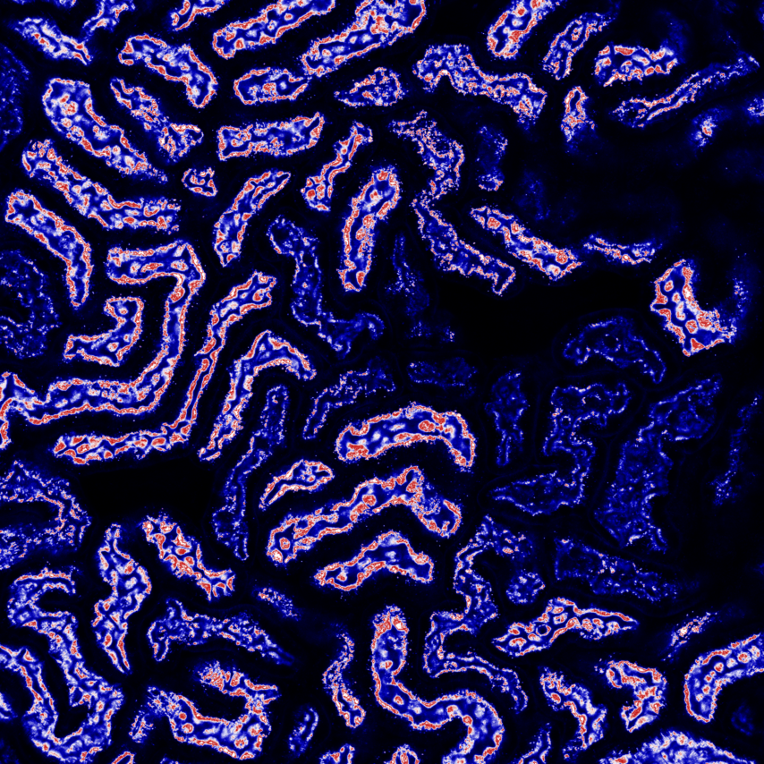
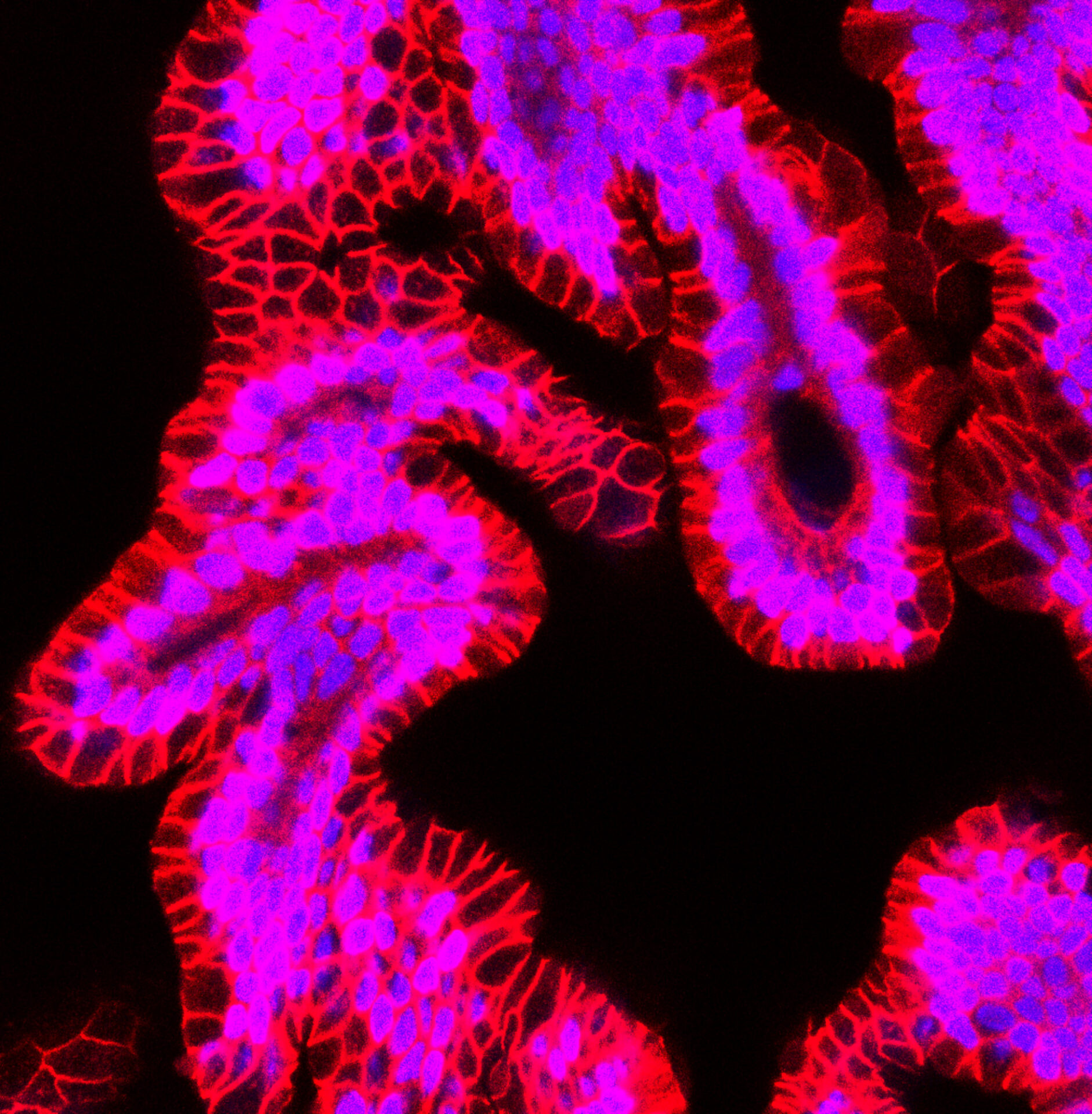
Adult human kidney organoids or tubuloids derived from CD24+ stem/progenitor cells. Red color in confocal image indicates E-cadherin (CDH1) staining, blue is DAPI.
From:
Yaoxian Xu and Rafael Kramann, Germany
Institute of Experimental Medicine and Systems Biology, Medical Clinic 2, Faculty of Medicine, RWTH Aachen University.
Author: Yaoxian Xu | Rafael Kramann, Germany
Cells
Info:
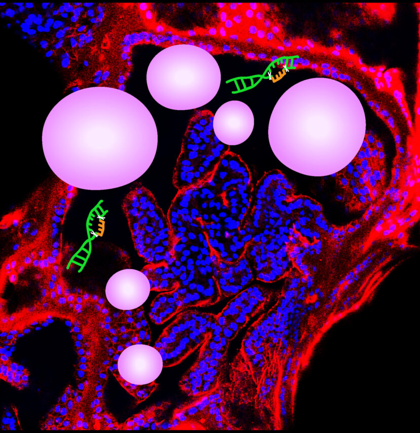
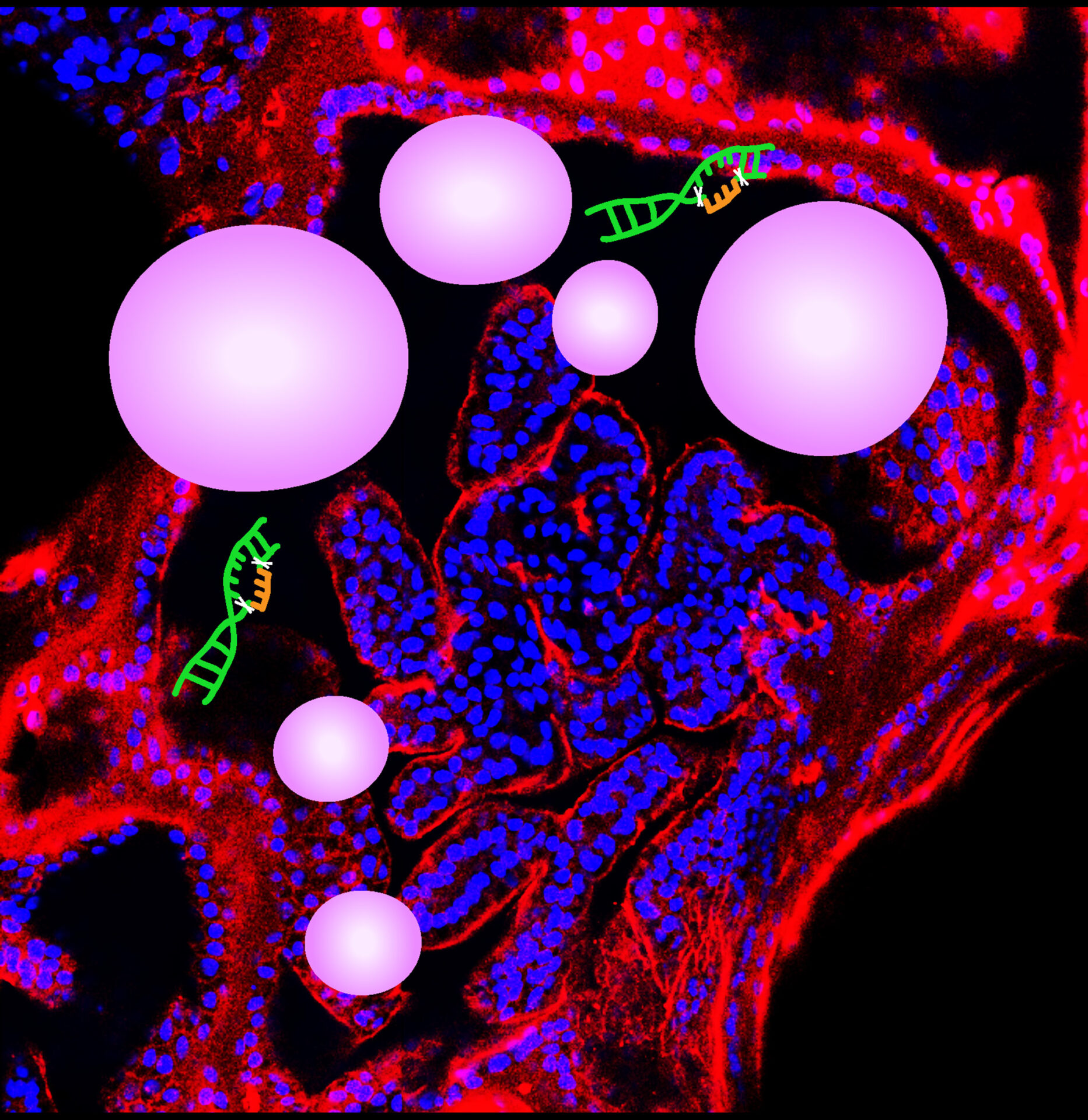
The Figure shows lenti-paired CRISPR-Cas9 gene editing for genetic disease ADPKD modeling in CD24+ derived adult human kidney tubuloids. Red color in Confocal image indicates CD24 staining, blue is DAPI, two cuts in DNA represent lenti-paired CRISPR-Cas9 gene editing in human PKD1 or PKD2 gene, white balloon-like structures represent ADPKD cysts.
From:
Yaoxian Xu and Rafael Kramann, Germany
Institute of Experimental Medicine and Systems Biology, Medical Clinic 2, Faculty of Medicine, RWTH Aachen University.
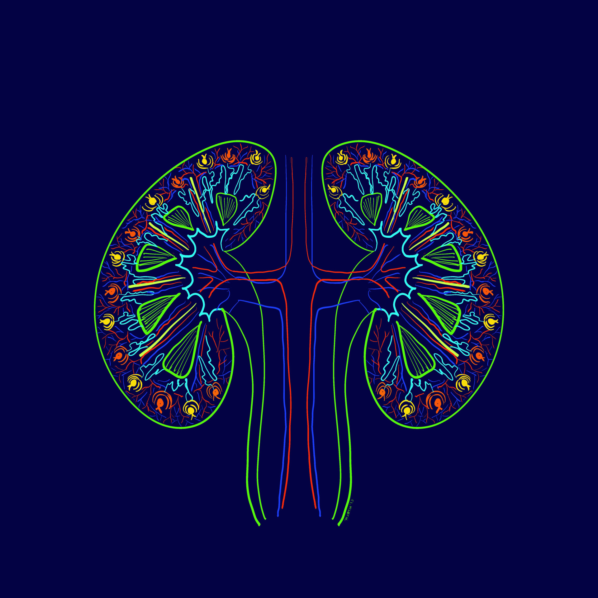
Author: Mayleen Laico, Philippines
Digital art
Info:
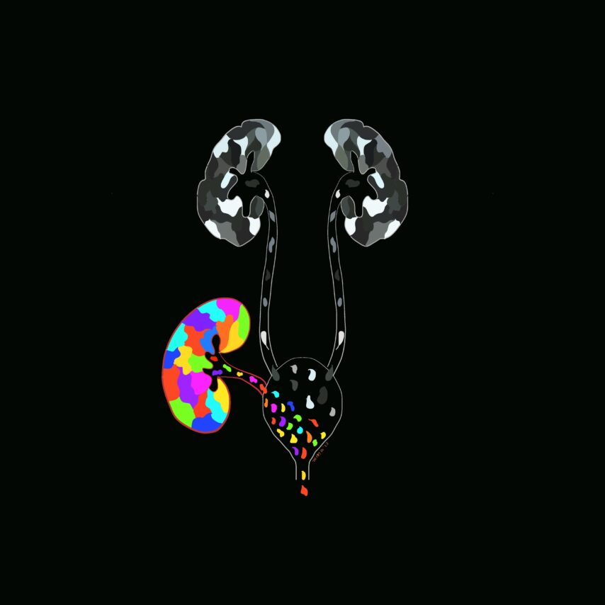
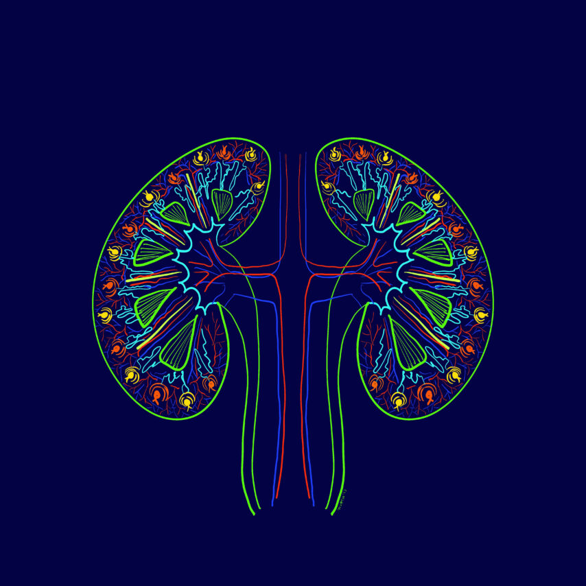
Organized Complexity shows the kidney’s complex function yet it is a very organized system
From:
Mayleen Laico, Philippines
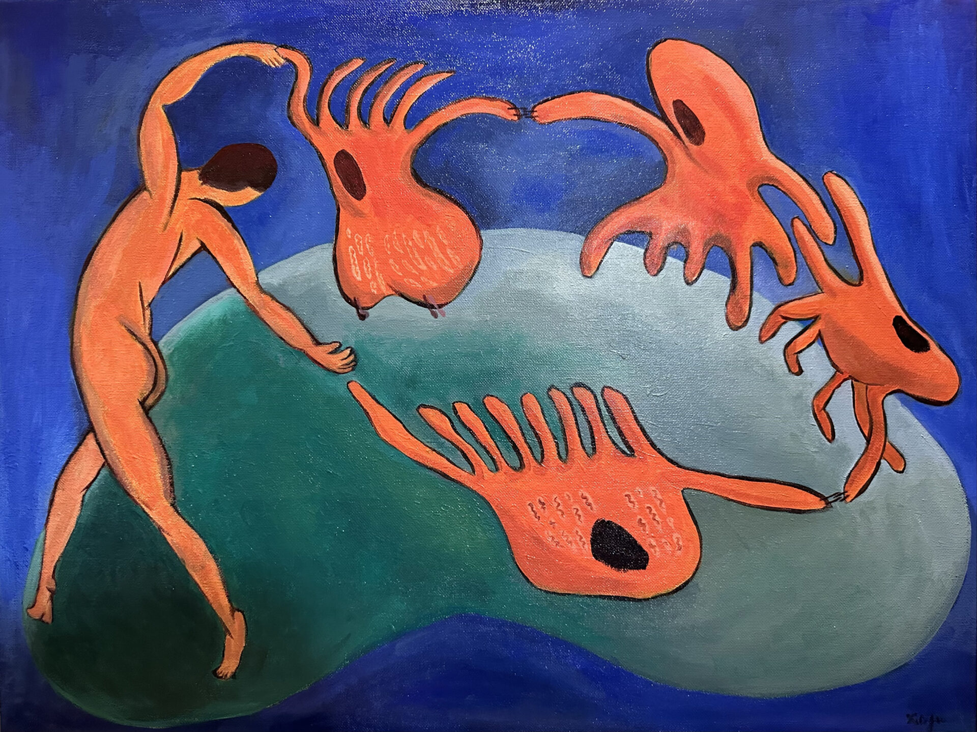
Author: Xinyu Dong, USA
Drawing
Info:
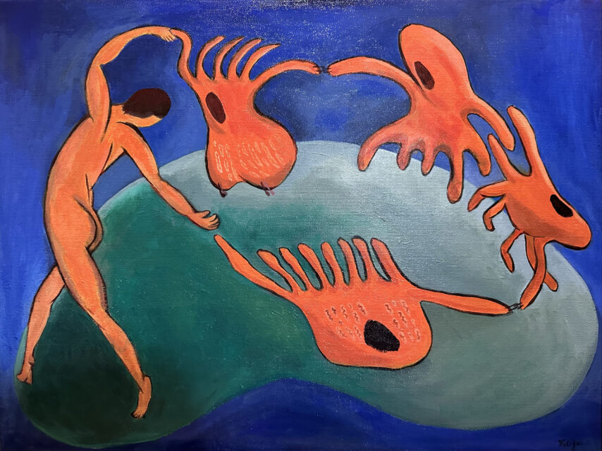
A nephrologist, immersed in passion, dances hand-in-hand with proximal tubule cells and podocytes. The dance unfolds on a green kidney, representing the earth, set against a blue cosmos background, symbolizing the nephrologist’s devotion to kidney research.
From:
Xinyu Dong, USA
Nephrology Department, Vanderbilt University Medical Center
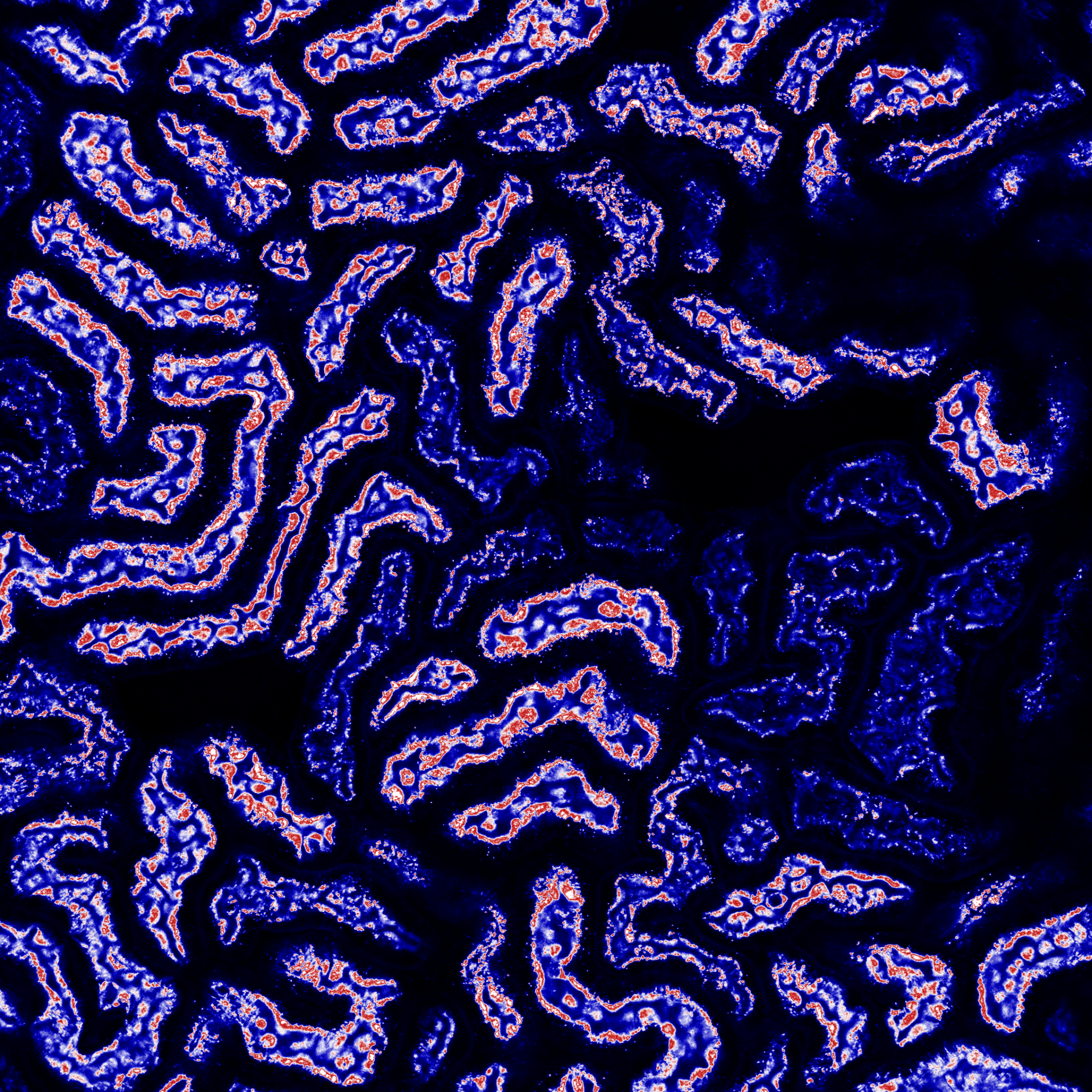
Author: Sara Gjeci | Meike N. Leiske | Adrian T. Press, Germany
Advanced microscopy
Info:
Drug delivery system targeting kidney
From:
Sara Gjeci, Meike N. Leiske, Adrian T. Press
University of Jena and Bayreuth, Germany

Author: Marieta Theodorakopoulou, Greece
Histology
Info:
Glomerulus Galaxy – Immunofluorescence images stained for C3 in a patient with lupus nephritis
From:
Marieta Theodorakopoulou, Greece
1st Department of Nephrology, Hippokration Hospital, Aristotle University of Thessaloniki,
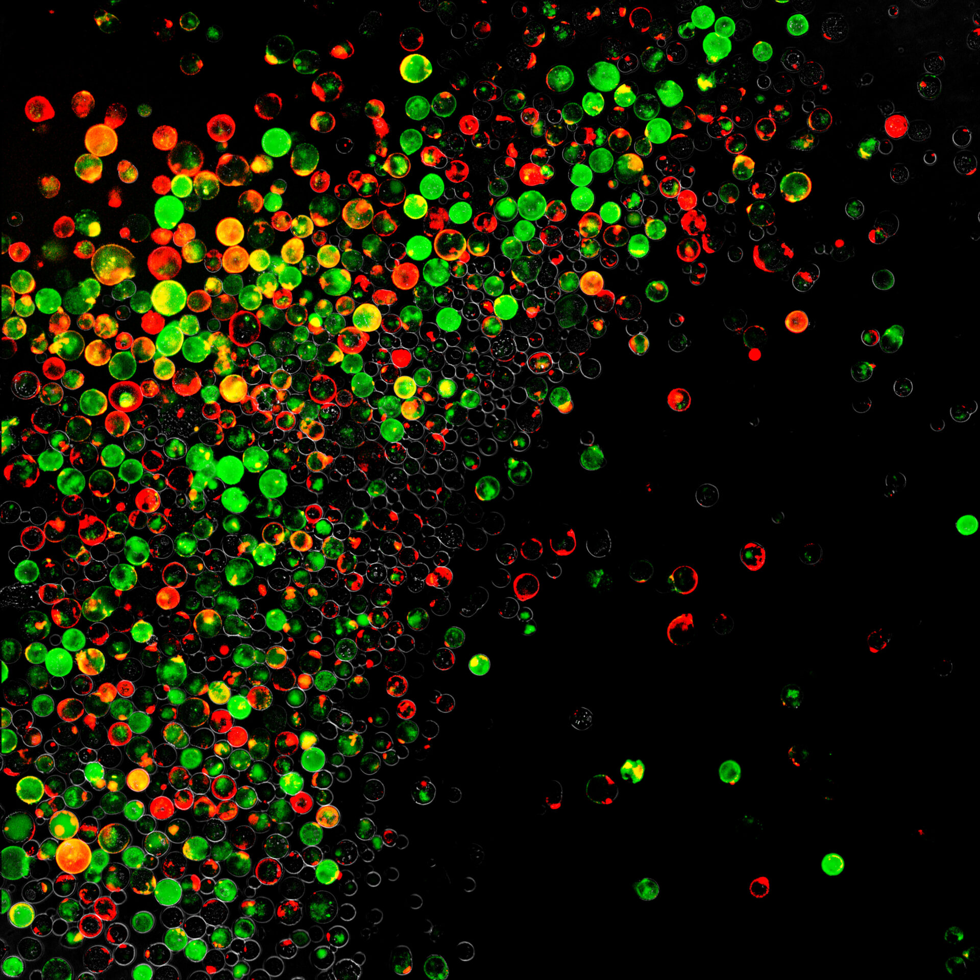
Author: Chenyu Li, Germany
Cells
NDT Cover December 2024
Info:
Immunofluorescence images of cultured urine stem cells (stained for CD133 (red) and CD24 (green), collected from healthy human urine.
From:
Chenyu Li, Germany
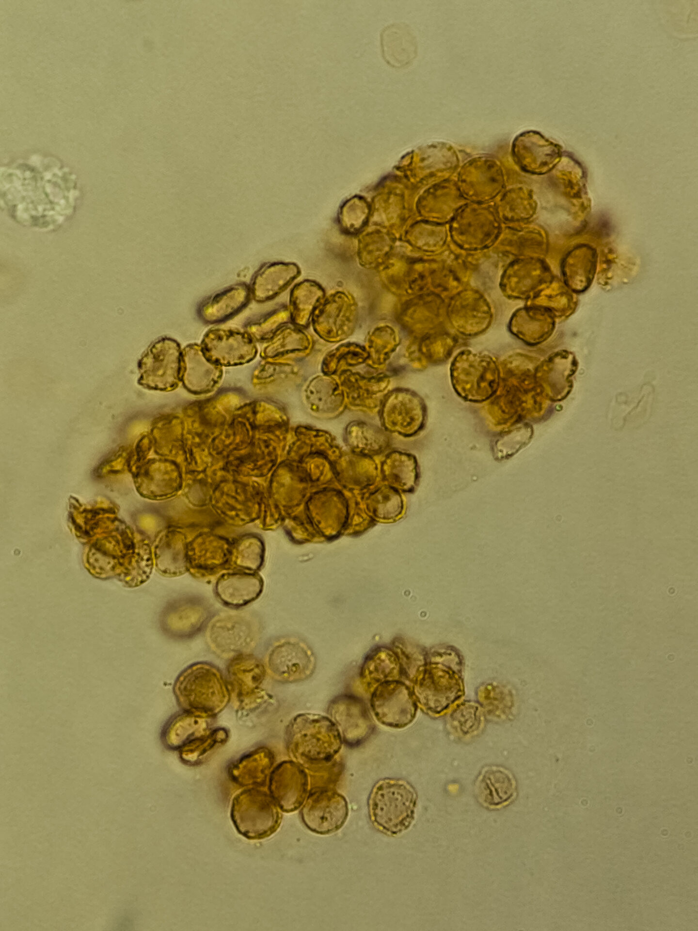
Author: Ralph Mohty, USA
Urine & urine microscopy
NDT Cover September 2024
Info:
Red blood cell cast indicating glomerulonephritis in a patient with AntiNeutrophil Cystoplasmic Antibody (ANCA) associated vasculitis
From:
Ralph Mohty, USA
MD MPH Stanford Nephrology
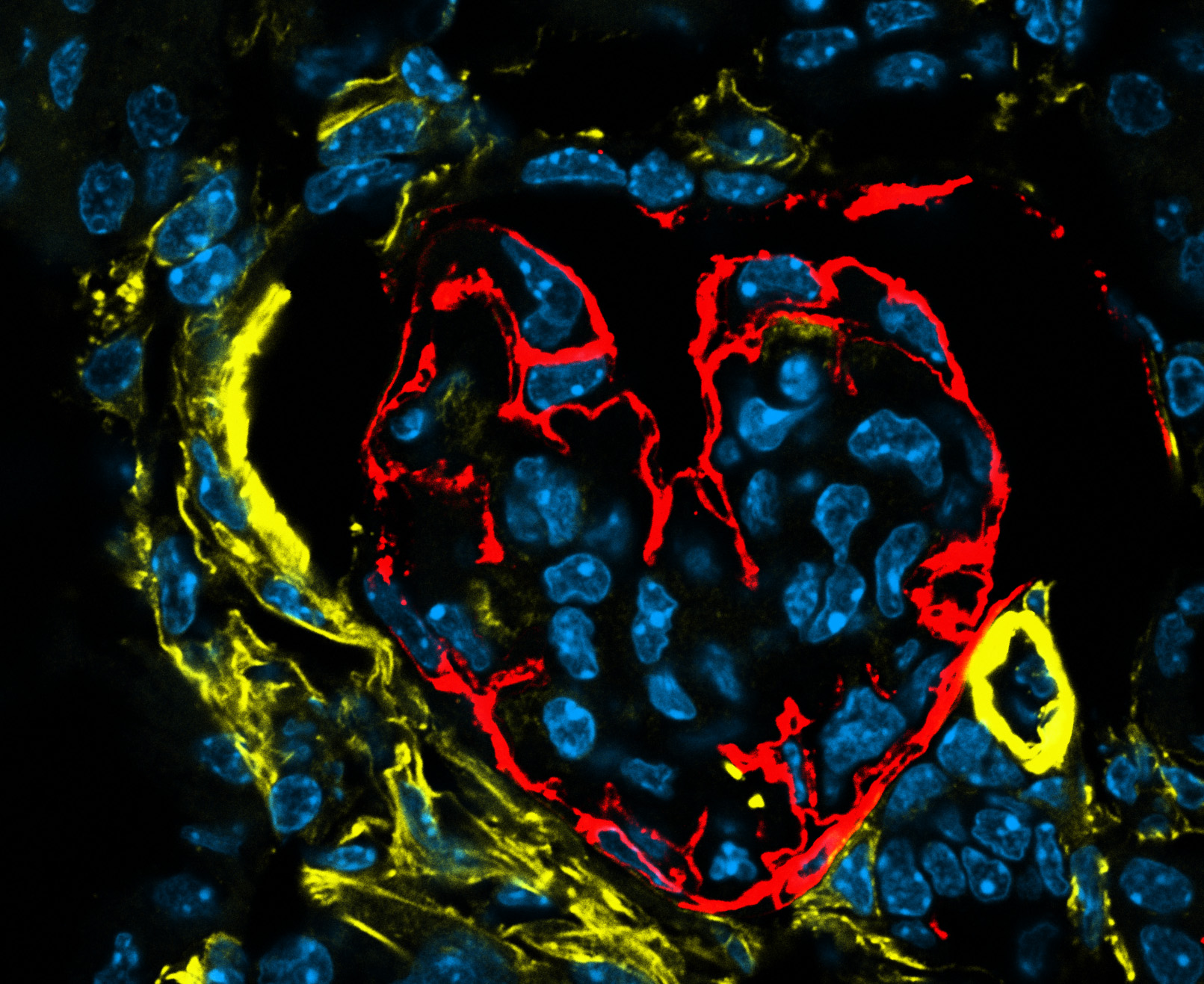
Author: Katharina Artinger, Austria
Advanced microscopy
NDT Cover February 2024
Info:
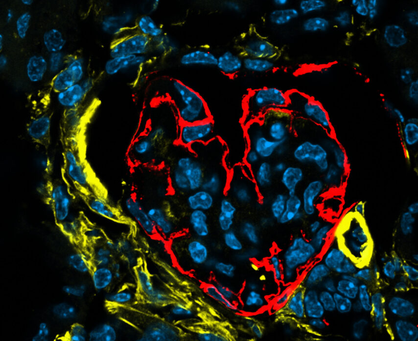
Love hurts – Heart-shaped glomerulus in a nephritic kidney
Podoplanin – red; Alpha smooth muscle actin – yellow; DAPI – blue.
From:
Katharina Artinger, Austria
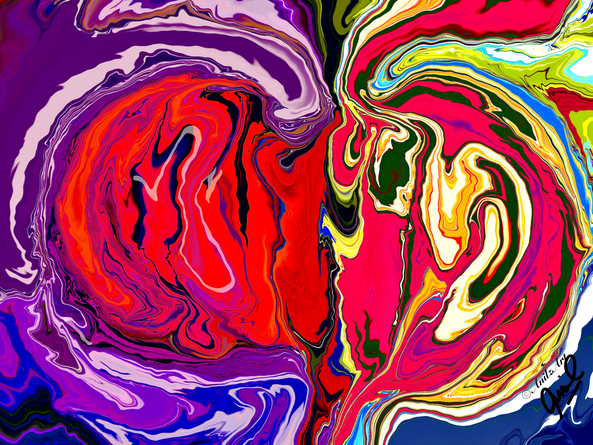
Author: Anil Kumar Saxena, United Arab Emirates
Digital art
NDT Cover July 2024
Info:
Focal Segmental Glomerulosclerosis
From:
Anil Kumar Saxena, United Arab Emirates
Chair, Nephrology Division Mediclinic Welcare Hospital Al Gahroud

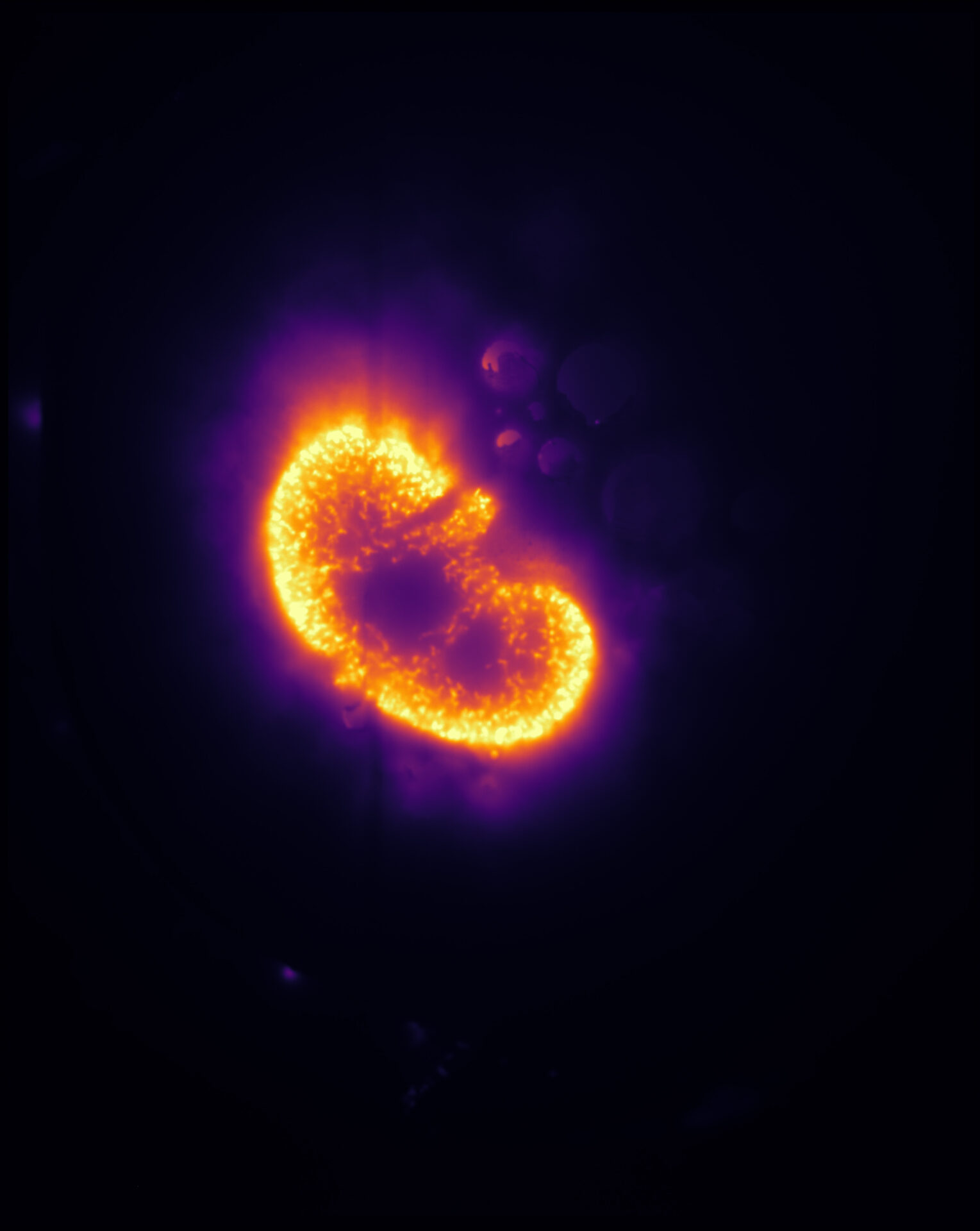
Author: Barbara Mara Klinkhammer, Germany
Histology
Info:
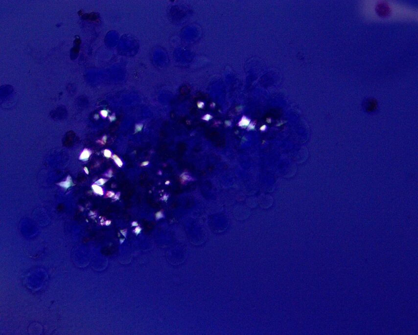
2,8-Dihydroxyadenine crystals in a rat kidney. PAS stain under polarized light.
From:
Barbara Mara Klinkhammer, Germany
Author: Sara Gjeci | Meike N. Leiske | Adrian T. Press, Germany
Imaging
Info:
Drug delivery system targeting kidney
From:
Sara Gjeci, Meike N. Leiske, Adrian T. Press
University of Jena and Bayreuth, Germany
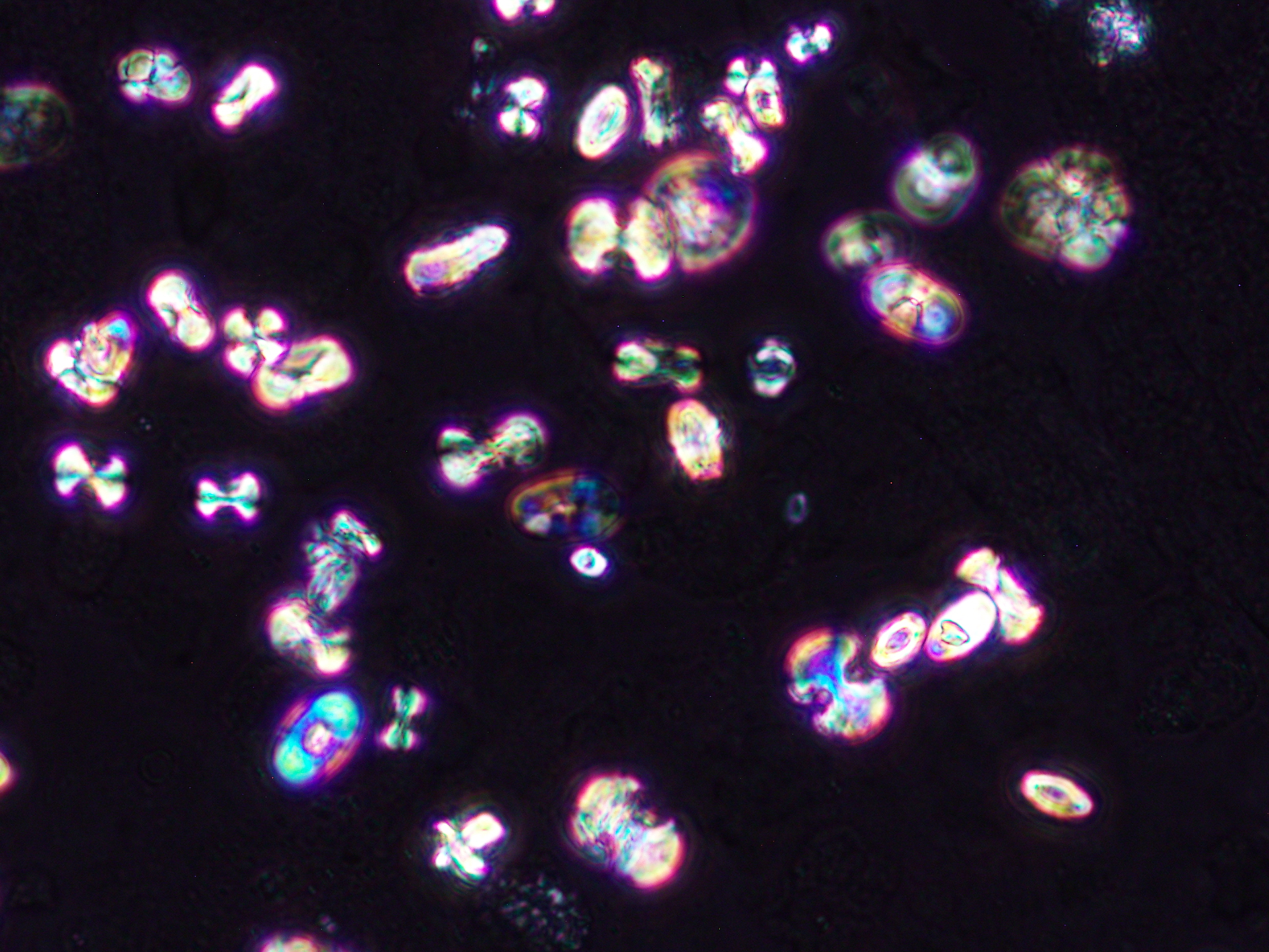
Author: Monarch Shah, USA
Urine & urine microscopy
Info:
Calcium oxalate monohydrate crystals in a patient with acute kidney injury, many of the dumbbell-shaped type seen under phase contrast and polarized light (Original magnification x400).
From:
Monarch Shah,
Fellow, University of Virginia, USA
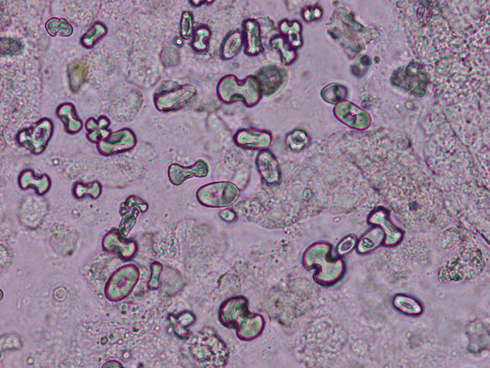
Author: Monarch Shah, USA
Urine & urine microscopy
Info:
Calcium oxalate monohydrate crystals in a patient with acute kidney injury, many of the dumbbell-shaped type seen under phase contrast and polarized light (Original magnification x400).
From:
Monarch Shah,
Fellow, University of Virginia, USA
Author: Ruben M Sandoval, USA
Advanced microscopy
Info:
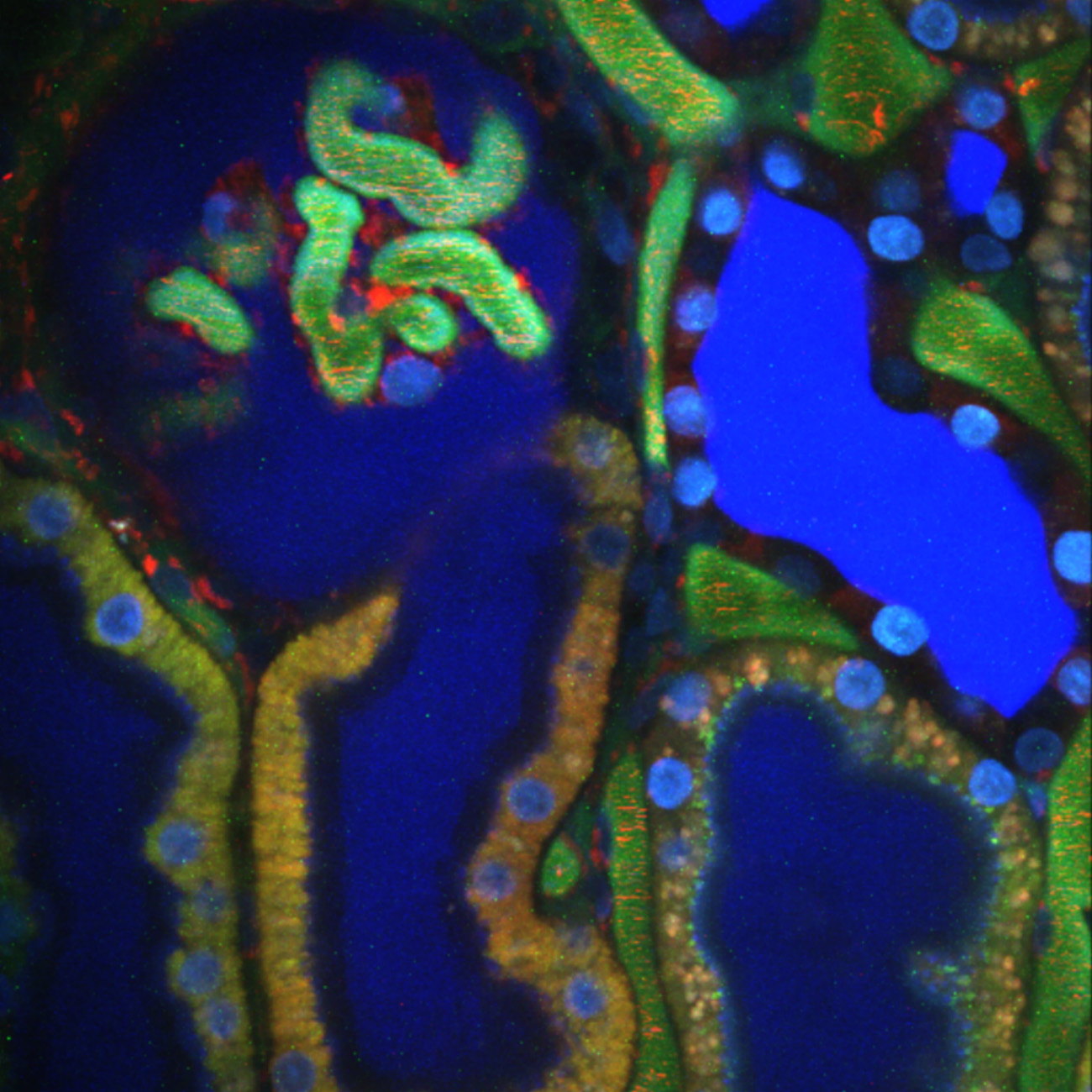
Time projection showing filtration on the surface of a rat kidney acquired using intravital 2-photon microscopy.
A bolus of a small blue dextran appears first in the lumen of the Bowman’s space, proximal tubule lumen, then finally in the cortical collecting duct lumen.
Projection is 300 frames, close to 4 minutes.
From:
Ruben M Sandoval, USA
Indiana Center for Biological Microscopy
Author: Ruben M Sandoval, USA
Advanced microscopy
NDT Cover December 2023
Info:
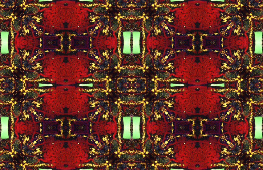
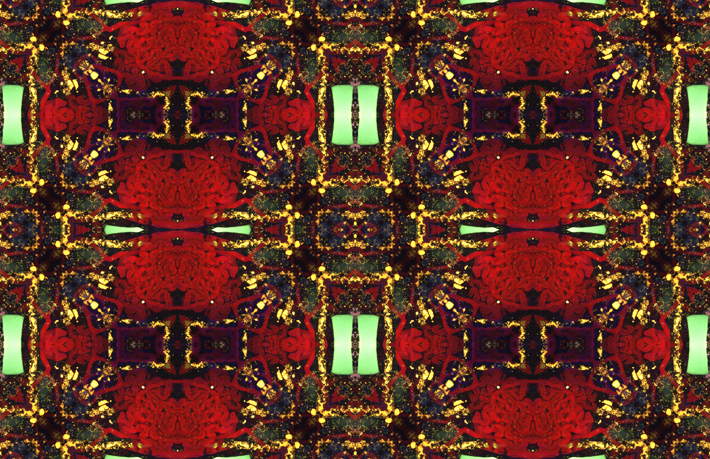
A chronic kidney disease rat model showing the distribution of albumin (red) acquired in 3D using intravital 2-photon microscopy.
Fluorescent cast material (green) can be seen obstructing the lumen of a distal tubule. The original image was processed to generate a tessellation with seamless borders and natural symmetry.
From:
Ruben M Sandoval, USA
Indiana Center for Biological Microscopy

Author: Hélène Fank, Belgium
Electron microscopy
Info:
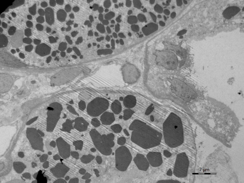
Electron microscopy confirmed the electron-dense crystalline structures within cytoplasm of proximal tubular epithelial cells
From:
Hélène Fank, Belgium
MD

Author: Hélène Fank, Belgium
Histology
Info:
Immunohistochemistry for kappa light chains was strongly positive within these crystals (b, white arrow)
From:
Hélène Fank, Belgium
MD
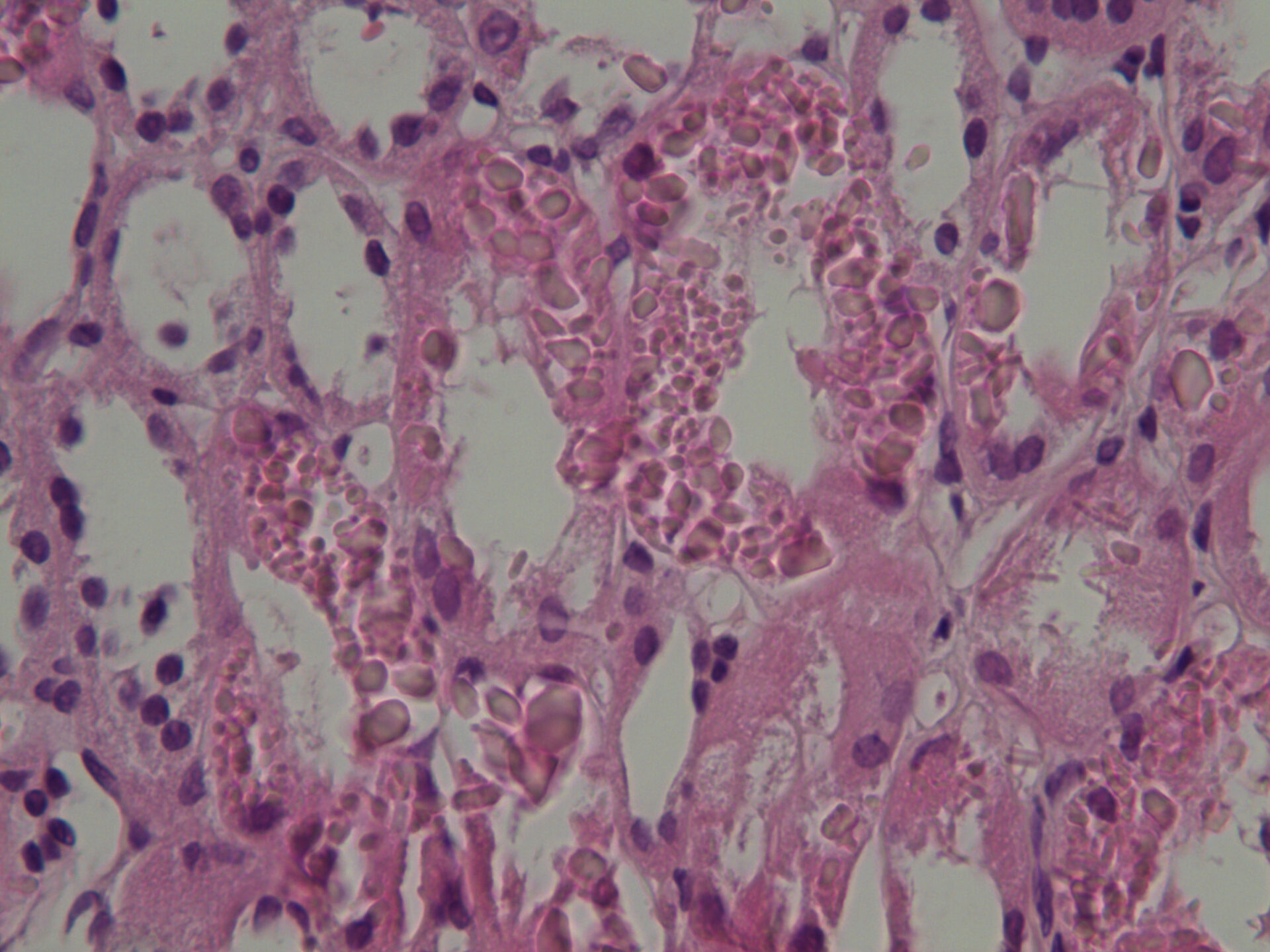
Author: Hélène Fank, Belgium
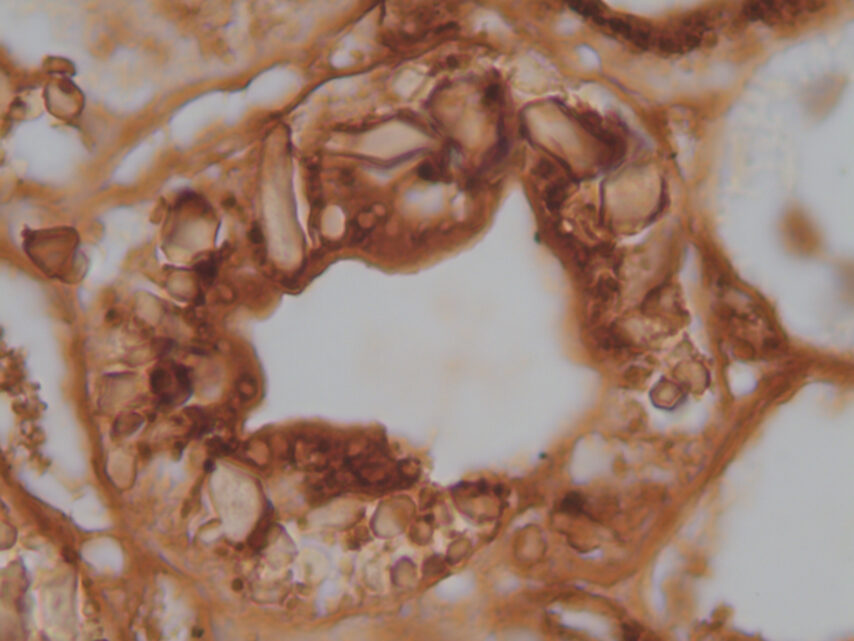
Histology
Info:
Light microscopy (Hematoxylin x400) showing intracellular crystalline inclusions within proximal tubular epithelial cells (a, black arrow)
From:
Hélène Fank, Belgium
MD

Author: Mayleen Laico, Philippines
Digital art
NDT Cover November 2023
Info:
The Greening of Nephrology
From:
Mayleen Laico, Philippines

Author: J. Romero Tafoya, México
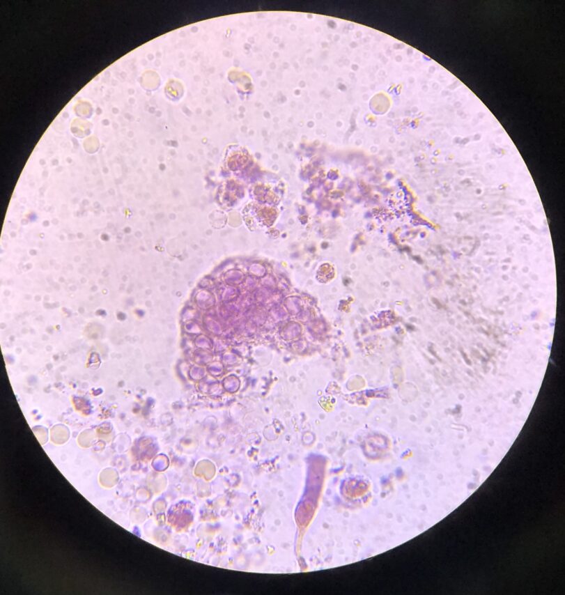
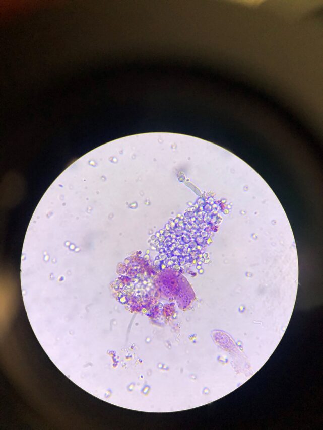
Urine & urine microscopy
Info:
Red blood cell cast – brightfield with Sternheimer – Malbin stain
From:
J. Romero Tafoya, México

Author: J. Romero Tafoya, México
Urine & urine microscopy
Info:
Bile cast and hyphae – brightfield with Sternheimer – Malbin stain
From:
J. Romero Tafoya, México
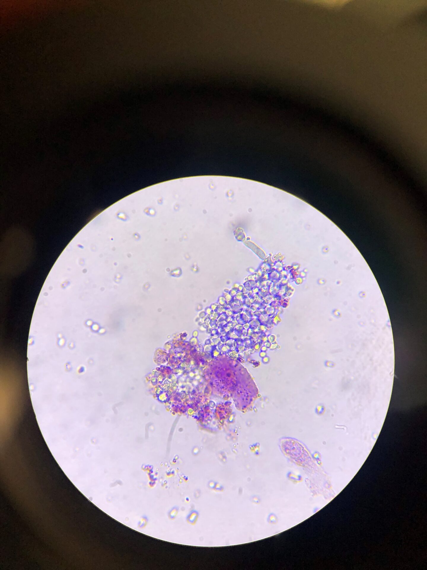
Author: J. Romero Tafoya, México
Urine & urine microscopy
Info:
Red blood cell cast- brightfield with Sternheimer- Malbin stain
From:
J. Romero Tafoya, México

Author: Anil Kumar Saxena
Digital art
Info:
Glomerulus – The ‘Heart’ of the Kidney
From:
Anil Kumar Saxena, United Arab Emirates
MD, FRCP, FASN Chair,
Nephrology Division Medical Director

Author: Reynaldo Noriega Flores, México
Drawing
Info:
Kidney Transplant
Watercolour painting
From:
Reynaldo Noriega Flores, México
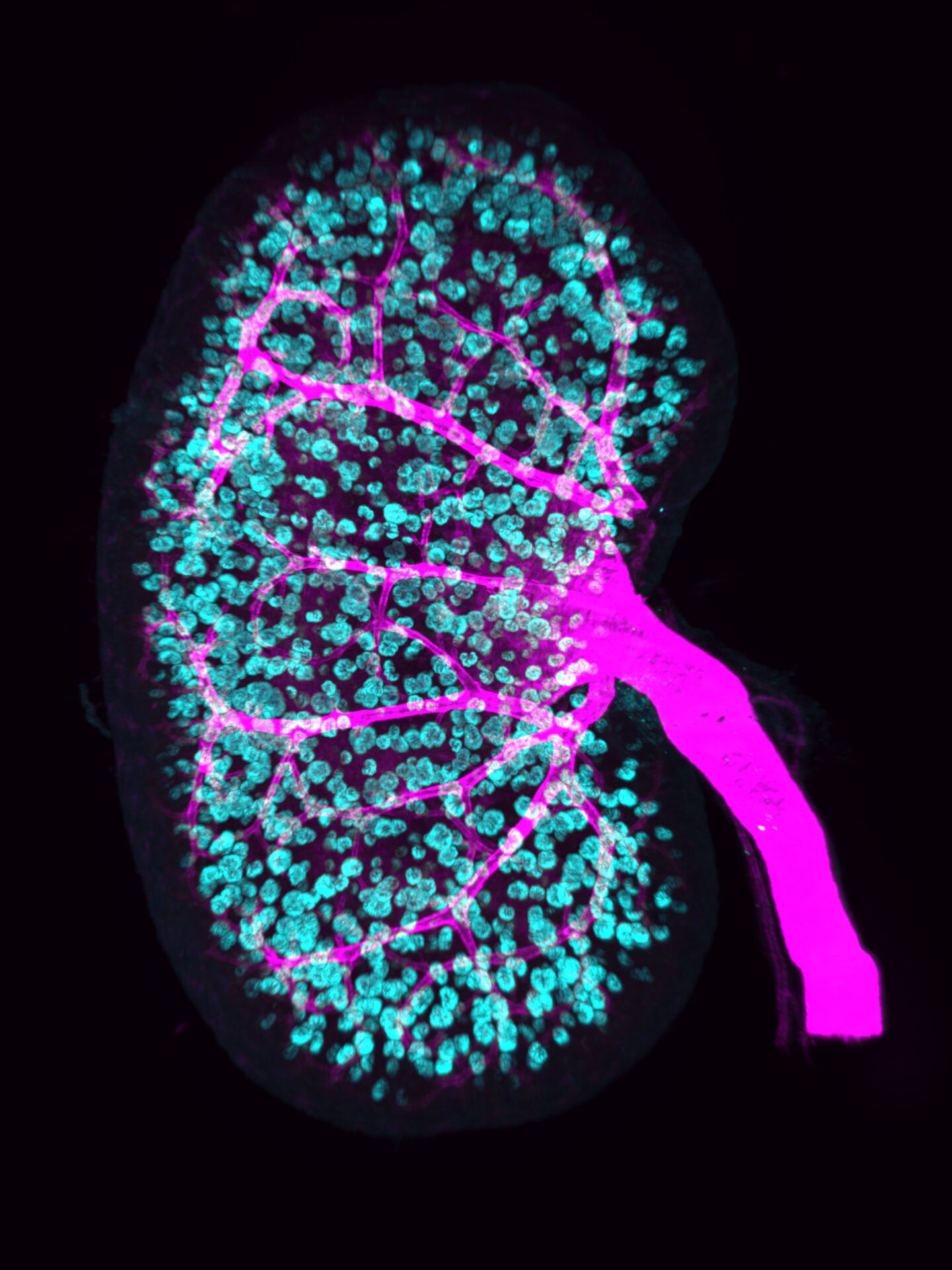
Author: Pierre-Emmanuel N'Guetta and Lori O'Brien, USA
Advanced microscopy
NDT Cover October 2023
Info:
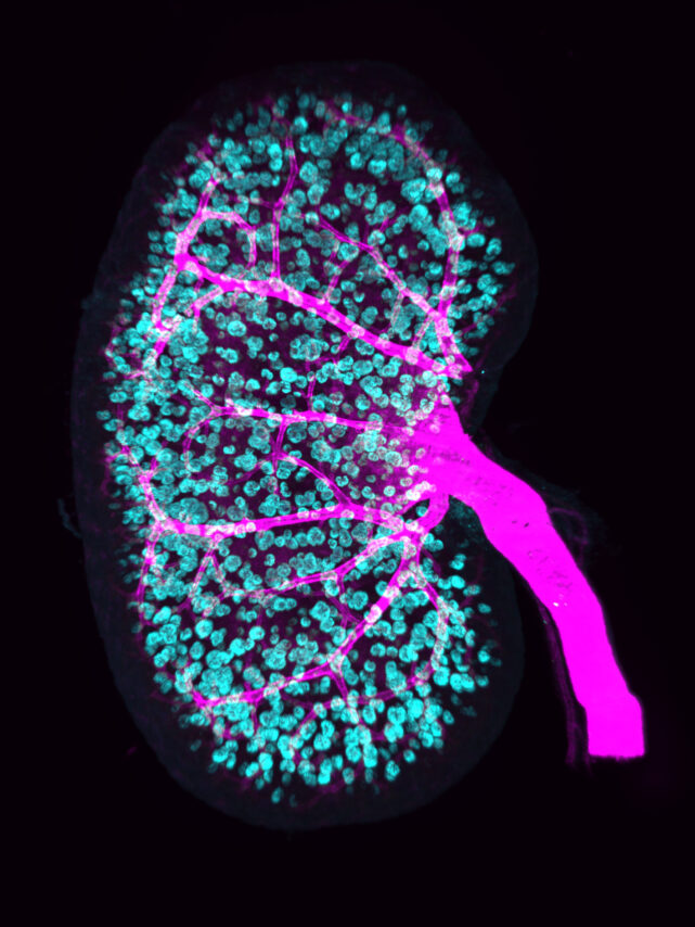
This image depicts a postnatal mouse kidney immunostained for the renal arterial tree with alpha-Smooth Muscle Actin (SMA, magenta) and glomeruli with podocin (Nphs2, cyan). Kidney was imaged with a LaVision Ultramicroscope II LightSheet using an Olympus MVPLAPO 2x/0.5 objective, a 2x Zoom, Sheet NA of 0.23, a Z step of 2.0µm and image pixel size of 1.54µm/pixel.
From:
Pierre-Emmanuel N’Guetta, USA
Lori O’Brien, USA
PhD, Assistant Professor
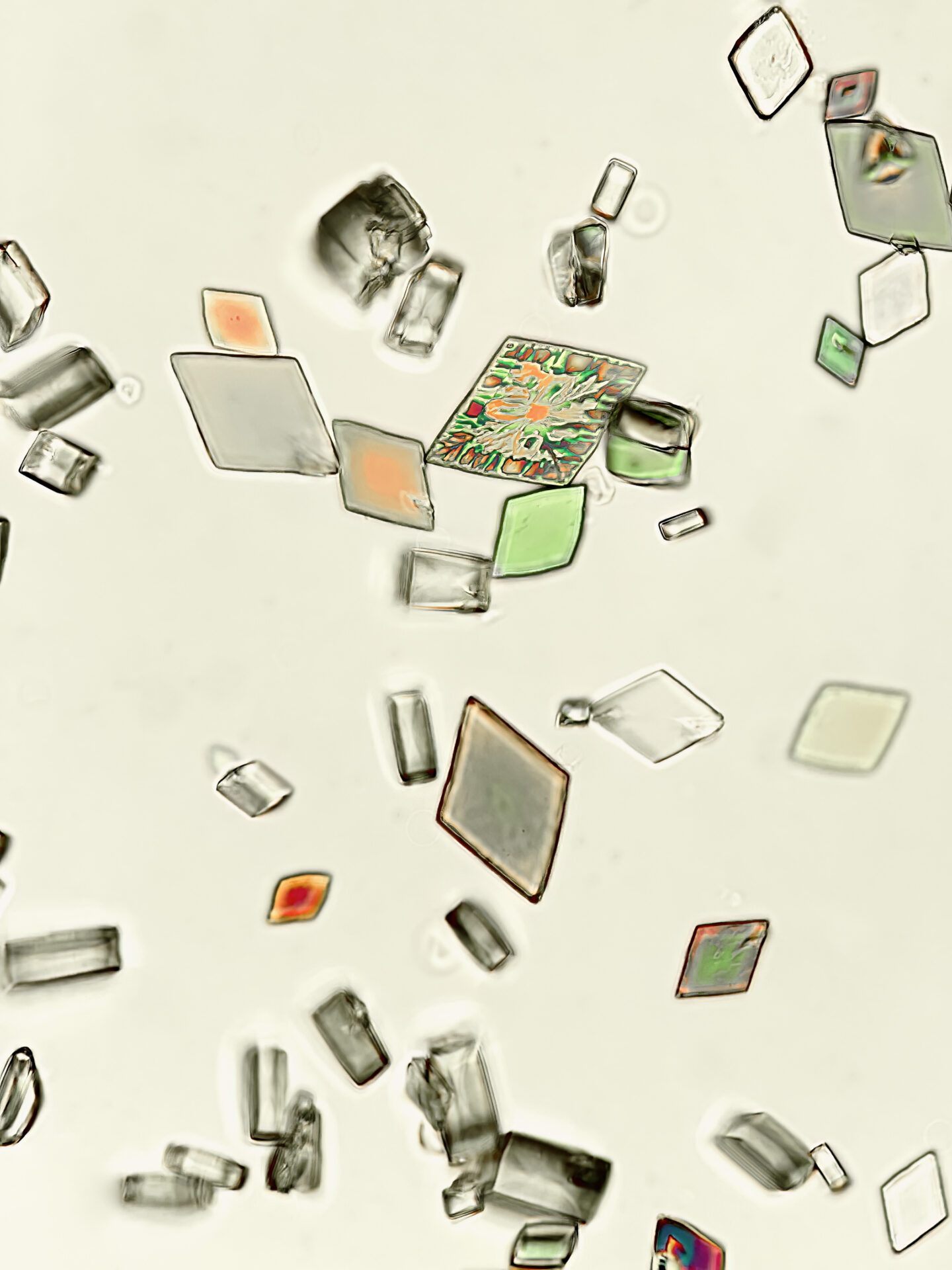
Author: Jay R. Seltzer, USA
Urine & urine microscopy
Info:
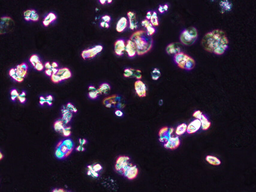
Uric acid crystals, under partial polarization – original magnification x400 – from patient with marked volume depletion and AKI
From:
Jay R. Seltzer, USA
MD, Chief of Nephrology

Author: Jay R. Seltzer, USA
Urine & urine microscopy
NDT March cover 2024
Info:
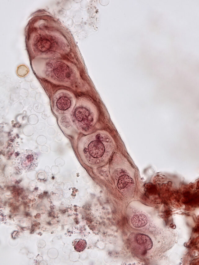
Renal tubular epithelial cell cast – brightfield with prolonged Sternheimer-Malbin staining – original magnification x1000 – from patient with bacterial endocarditis, acute kidney injury, and infection related glomerulonephritis
From:
Jay R. Seltzer, USA
MD, Chief of Nephrology

Author: Jay R. Seltzer, USA
Urine & urine microscopy
NDT Cover September 2023
Info:
Uric acid crystals in urine, under fully polarized light – original magnification x400 – from patient with marked volume depletion and AKI
From:
Jay R. Seltzer, USA
MD, Chief of Nephrology
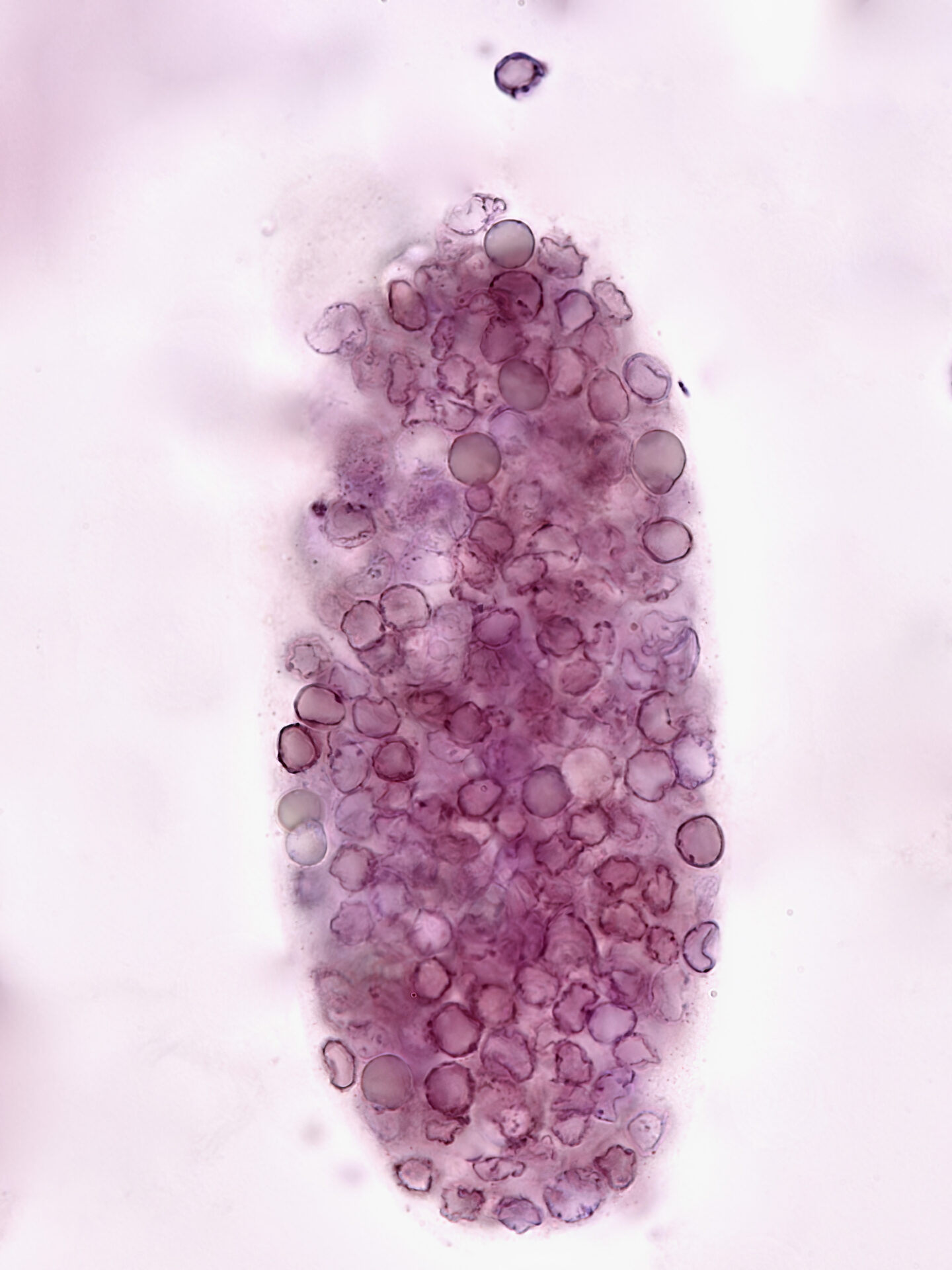
Author: Jay R. Seltzer, USA
Urine & urine microscopy
Info:
Red blood cell cast – brightfield with Sternheimer-Malbin stain – original magnification x1000 – from patient with hydralazine-induced vasculitis
From:
Jay R. Seltzer, USA
MD, Chief of Nephrology

Author: Tiffany Caza, USA
Drawing
NDT Cover July 2023
Info:
This image demonstrates an abstract view of the transition between a proximal tubule and the thick descending limb of the loop of Henle
From:
Tiffany Caza, USA
MD/PhD, a nephropathologist at Arkana Laboratories

Author: Swetha Kanduri, USA
Urine & urine microscopy
Info:
B: Bright field, dark field and phase contrast image of cluster of calcium oxalate dihydrate crystals surrounding leukocytes
From:
Swetha Kanduri, USA
MD, Assistant Professor of Medicine
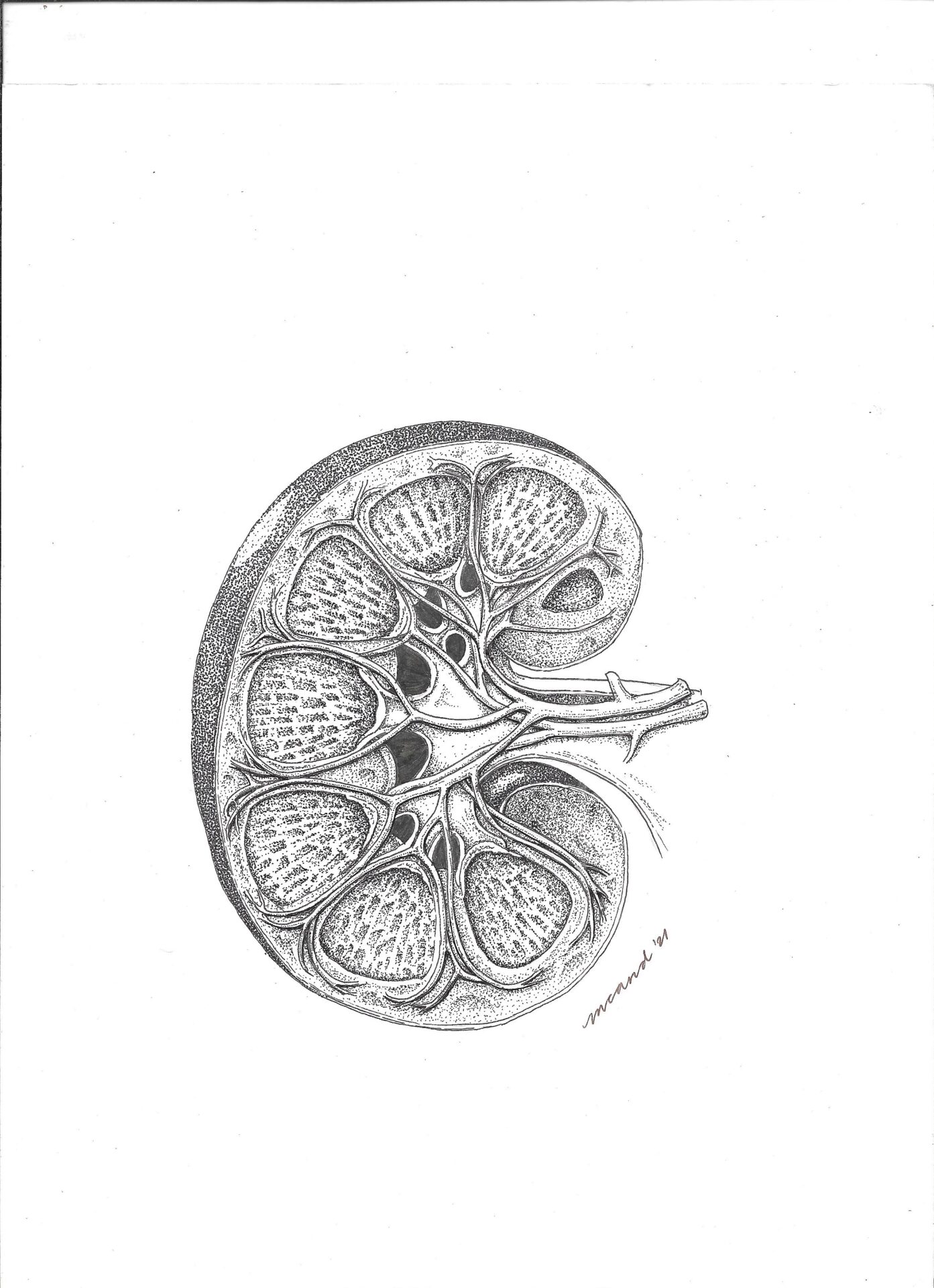
Author: Maia Celeste Arbatin, Philippines
Drawing
Info:
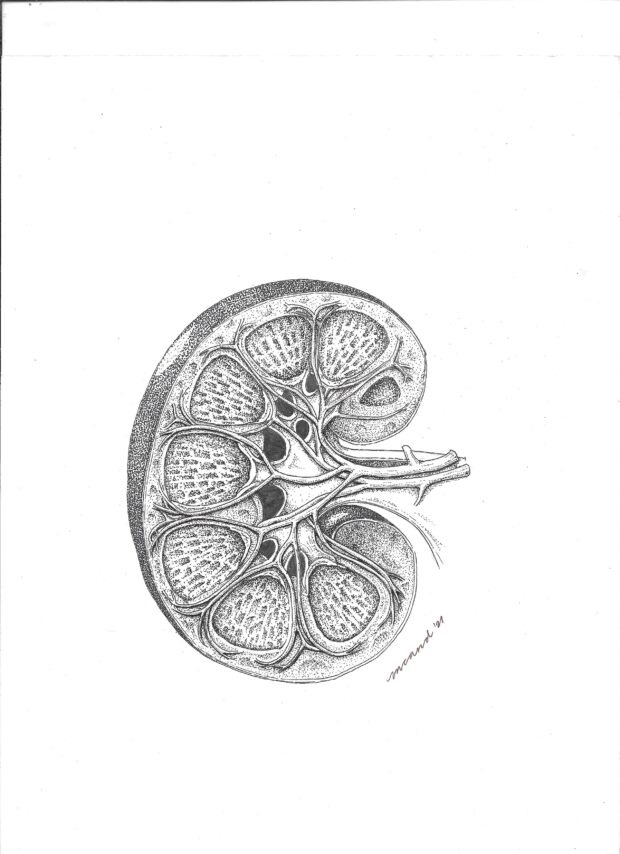
Pen and ink sketch
From:
Maia Celeste Arbatin, Philippines
MD FPCP FPSN (#nephgirlsketches)

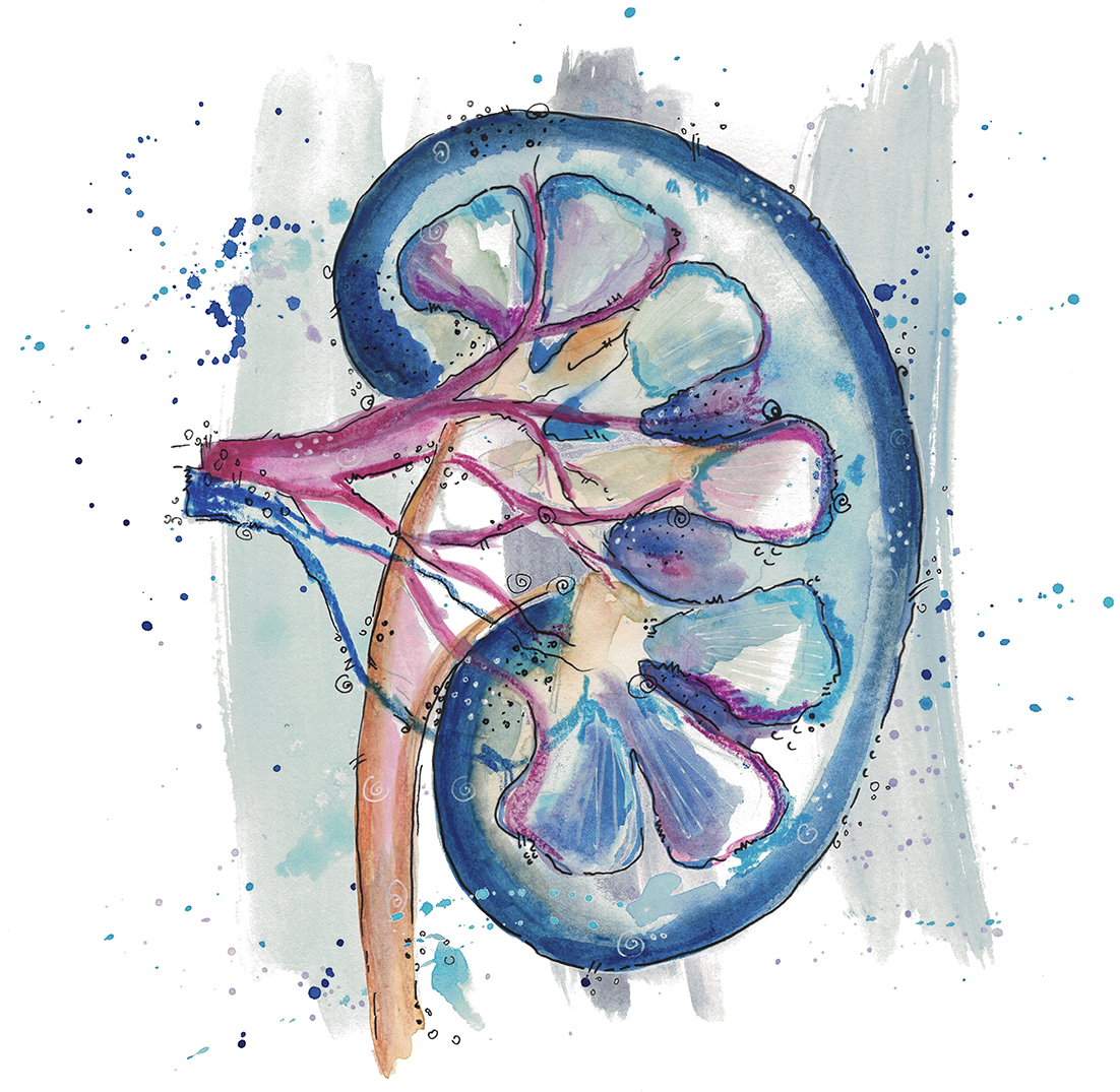
Author: Maia Celeste Arbatin, Philippines
Drawing
Info:
Hydrangea Kidney
From:
Maia Celeste Arbatin, Philippines
MD FPCP FPSN (#nephgirlsketches)
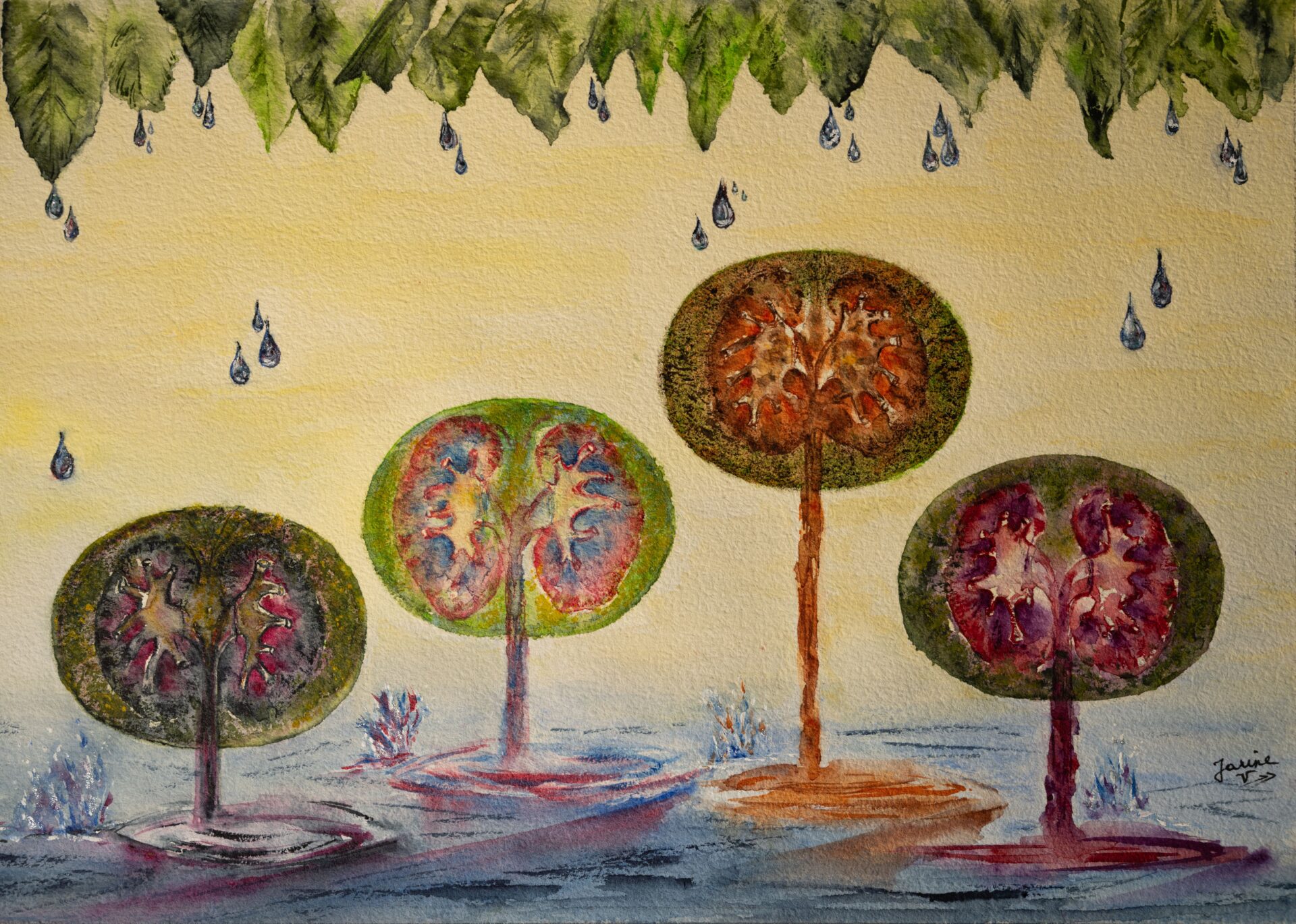
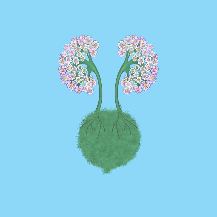
Author: Jeanine Verbeke, Belgium
Drawing
Info:
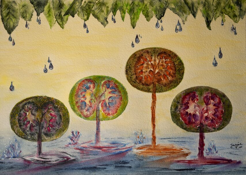
Kidney Trees
From:
Jeanine Verbeke, Belgium
Author: Dominique Couck, Belgium
Drawing
NDT Cover August 2023
Info:
Drawing for a family gift
From:
Dominique Couck, Belgium

Author: Boonyarit Cheunsuchon, Thailand
Histology
Info:
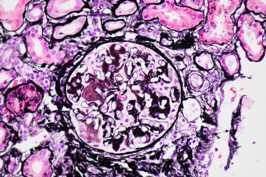
Eyelash sign in glomerular amyloidosis
From:
Boonyarit Cheunsuchon, Thailand
MD, Associate professor

Author: Sonja Djudjaj, Germany
Digital art
Info:
NDT history
From:
Sonja Djudjaj, Germany
PhD, Assistant Professor

Author: Sonja Djudjaj, Germany
Drawing
Info:
Water color kidney
From:
Sonja Djudjaj, Germany
PhD, Assistant Professor
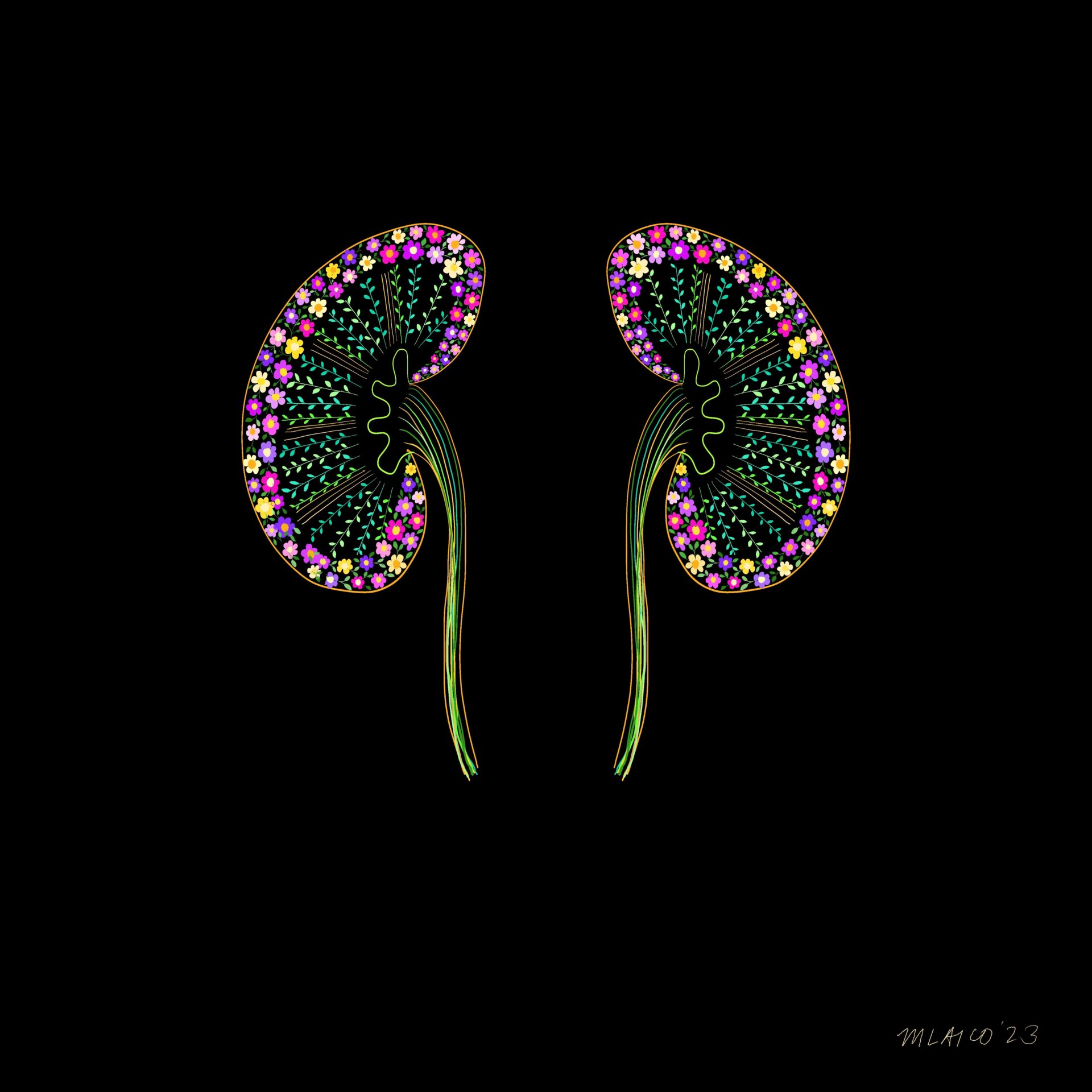
Author: Mayleen Laico, Philippines
Digital art
Info:
Giving hope
From:
Mayleen Laico, Philippines
MD

Author: Maria Lucia Angelotti, Italy
Advanced microscopy
Info:
Representative image obtained by STED super-resolution microscopy, showing strong positivity for IgA deposits (orange) and a partial mild effacement of foot processes (stained with nephrin, cyan) in a biopsy of IgA nephropathy patient. The biopsy specimen underwent optical tissue clearing before staining.
From:
Maria Lucia Angelotti, Italy
PhD
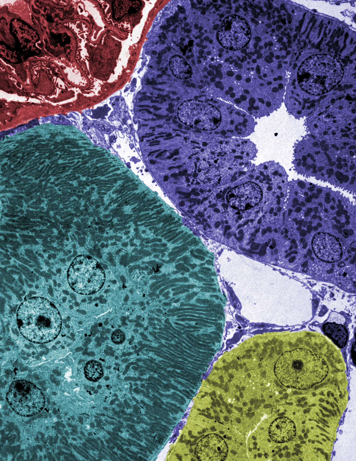
Author: Miriam Buhl, Germany
Electron microscopy
Info:
Colorized transmission electron microscopy image of murine renal kidney cortex. Individual tubular segments are colored in green, blue and yellow, while a glomerular section is colored in red.
From:
Miriam Buhl, Germany
PhD
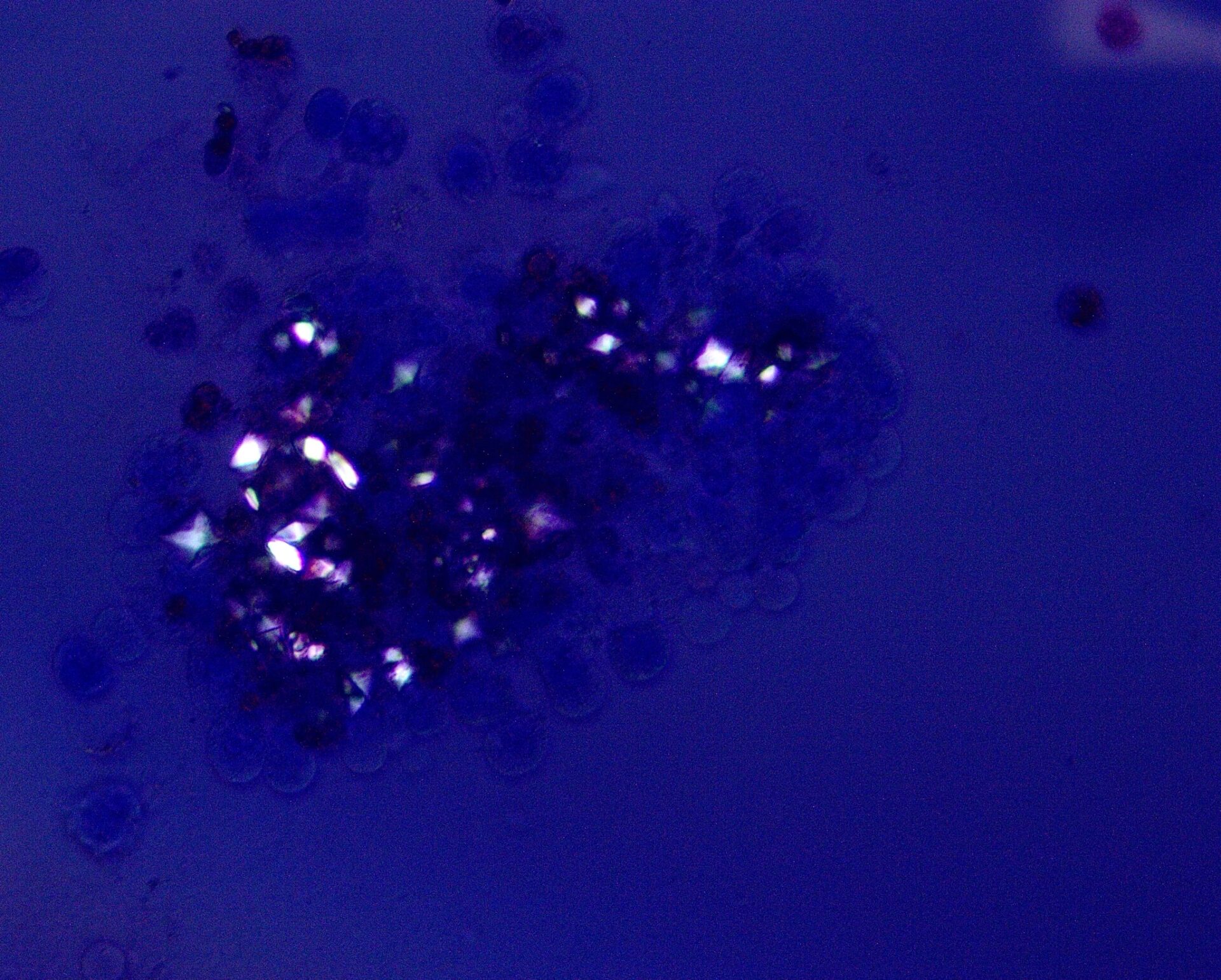
Author: Swetha Kanduri, USA
Urine & urine microscopy
Info:
Phase contrast image of cluster of calcium oxalate dihydrate crystals surrounding leukocytes
From:
Swetha Kanduri, USA
MD, Assistant Professor of Medicine
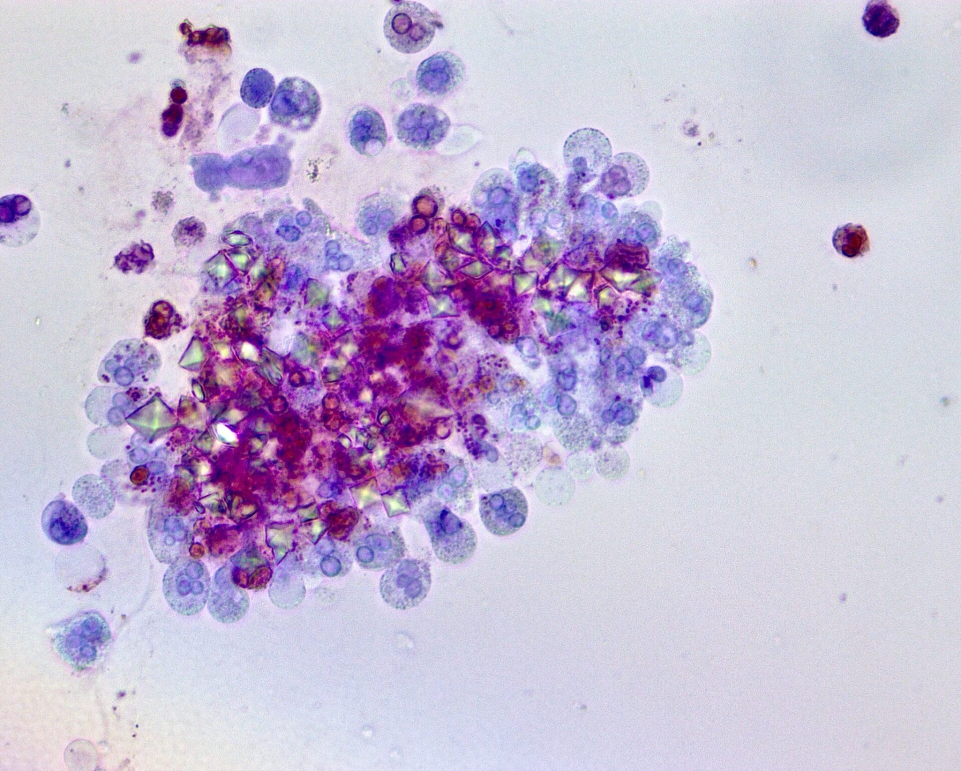
Author: Swetha Kanduri, USA
Urine & urine microscopy
Info:
Bright field image of cluster of calcium oxalate dihydrate crystals surrounding leukocytes
From:
Swetha Kanduri, USA
MD, Assistant Professor of Medicine
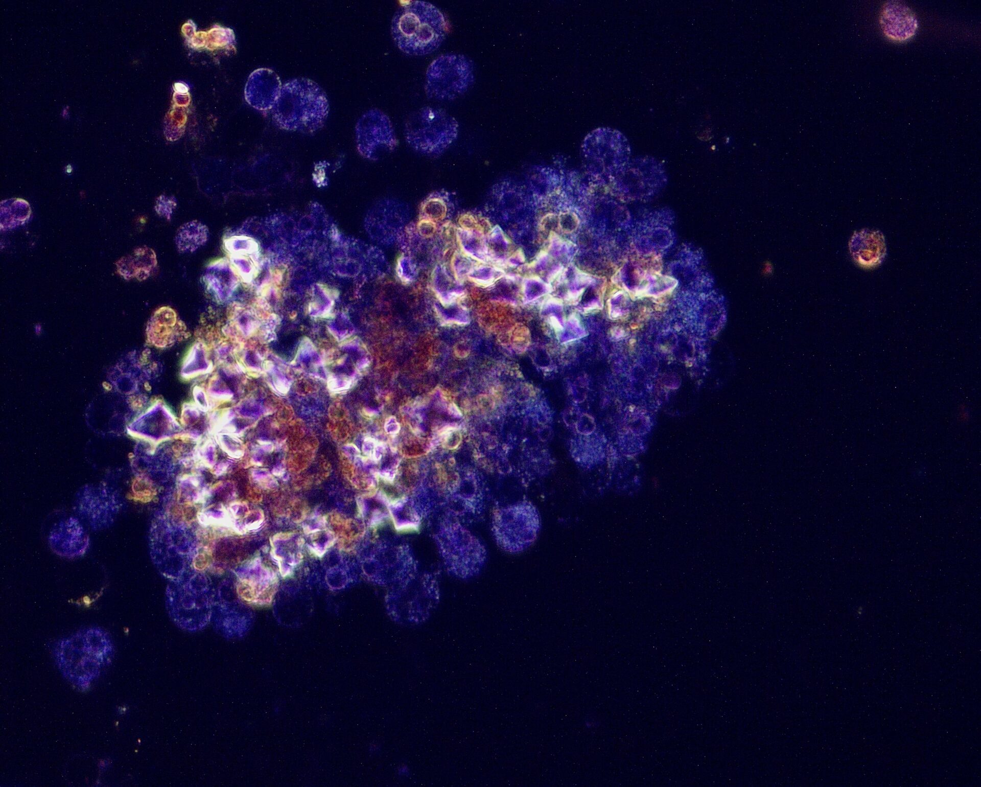
Author: Swetha Kanduri, USA
Urine & urine microscopy
Info:
Dark field image of cluster of calcium oxalate dihydrate crystals surrounding leukocytes
From:
Swetha Kanduri, USA
MD, Assistant Professor of Medicine

Author: Jay R. Seltzer, USA
Urine & urine microscopy
Info:
Red blood cell cast – brightfield with Sternheimer-Malbin stain – original magnification x1000 – from patient with anti-glomerular basement membrane disease
From:
Jay R. Seltzer, USA
MD, Chief of Nephrology
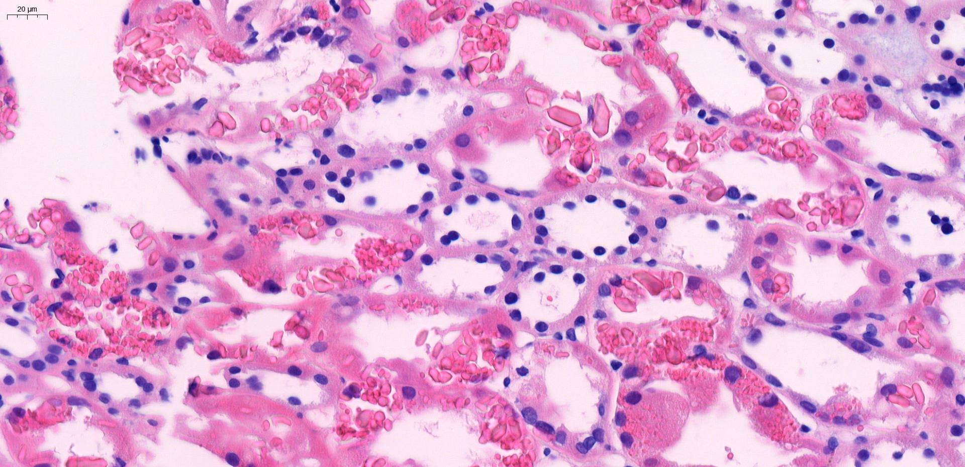
Author: Hélène Fank, Belgium
Histology
Info:
Secondary Toni-Debré-FANCONI syndrome – intracellular crystalline inclusions within proximal tubular epithelial cells
From:
Hélène Fank, Belgium
MD
Co-authors:
Antoine Bouquegneau, Christophe Bovy and Orphal Colleye
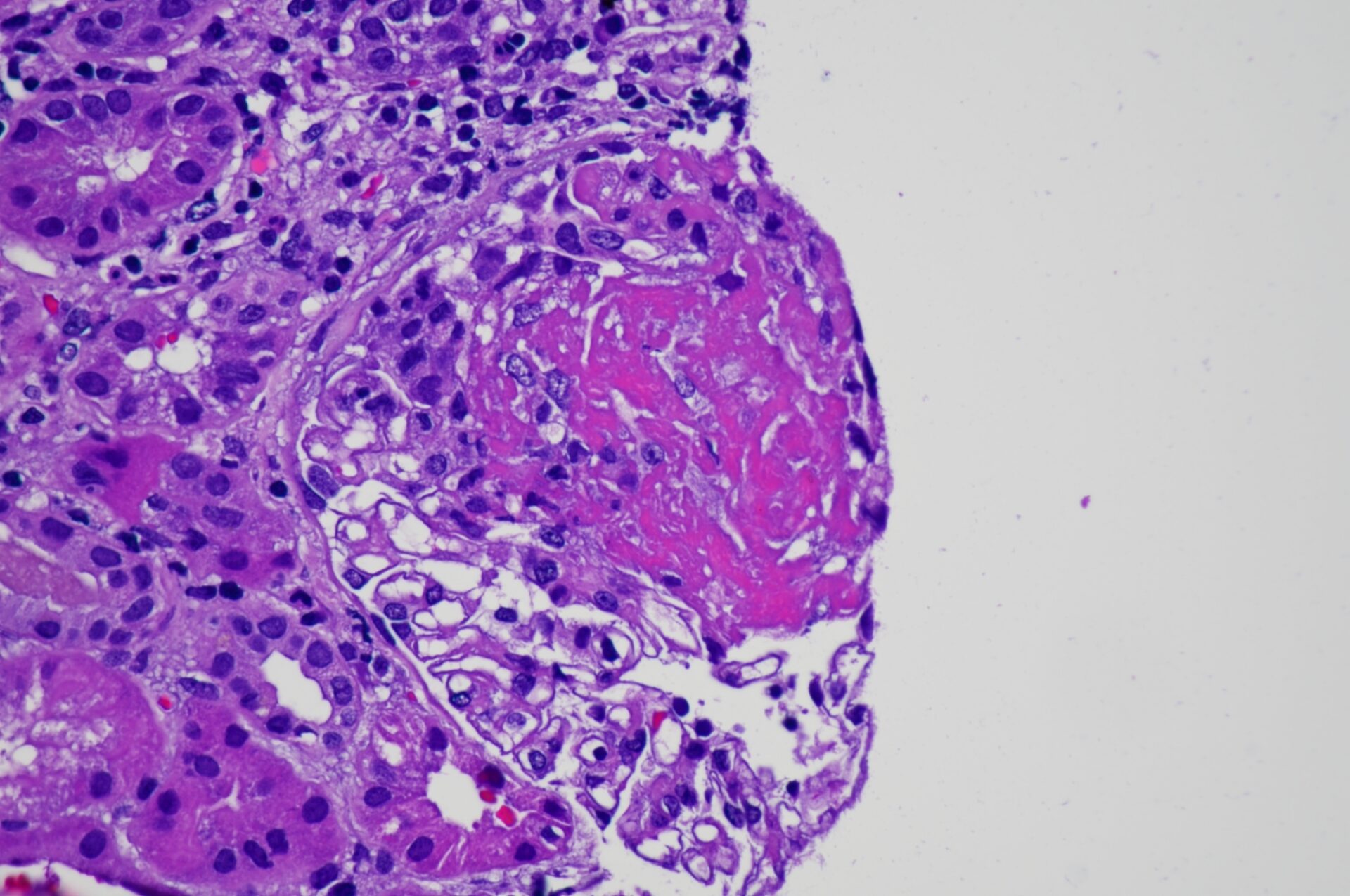
Author: Boonyarit Cheunsuchon, Thailand
Histology
Info:
Fibrinoid necrosis in a patient with ANCA-associated glomerulonephritis
From:
Boonyarit Cheunsuchon, Thailand
MD, Associate professor
Acknowledgements
NDT Kidney Art Editor:
Barbara Mara Klinkhammer, Germany
Also interested in submitting an image to NDT? Please contact ndt@era-online.org


